
IMAGE
Fig. 9
- ID
- ZDB-IMAGE-221127-9
- Publication
- Webster et al., 2021 - Role of BMP signaling during early development of the annelid Capitella teleta
- All Figures
- Figures for Webster et al., 2021
Image
|
Figure Caption
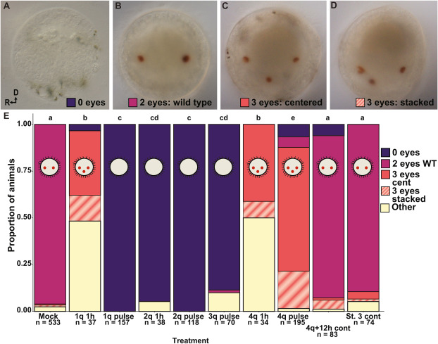
Fig. 9 Fig. 9. Larval eye pigment cells after BMP treatment. A–D Most common eye pigment cell (orange) phenotypes, anterior view. A) 0 eyes (1q pulse), B) 2 wild-type eyes (1q pulse), C) 3 eyes centered (1q pulse), D) 3 eyes stacked (1q pulse). R, right side; D, dorsal. E. Proportion of animals with each eye phenotype in each BMP treatment (colors correspond to boxes in A–D, and yellow: other (see text for a description of other eye phenotypes). Letters indicate significance groups.
Acknowledgments
This image is the copyrighted work of the attributed author or publisher, and
ZFIN has permission only to display this image to its users.
Additional permissions should be obtained from the applicable author or publisher of the image.
Reprinted from Developmental Biology, 478, Webster, N.B., Corbet, M., Sur, A., Meyer, N.P., Role of BMP signaling during early development of the annelid Capitella teleta, 183-204, Copyright (2021) with permission from Elsevier. Full text @ Dev. Biol.