
Fig. 12
- ID
- ZDB-IMAGE-221127-12
- Publication
- Webster et al., 2021 - Role of BMP signaling during early development of the annelid Capitella teleta
- All Figures
- Figures for Webster et al., 2021
|
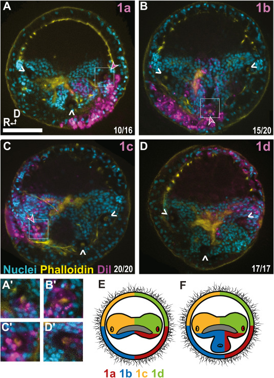
Fig. 12 Fig. 12. DiI localization and fate map of the eyes and brain of animals treated with BMP with a 4q pulse (Z-stack). A) 1a injected, showing contributions to the left brain lobe and eye B) 1b injected, showing contributions to the right brain lobe and the ectopic eye and brain lobe C) 1c injected, showing contributions to the right brain lobe. D) 1d injected, showing contributions to the left brain lobe. A′-C′) Inset of eye with DiI labeling (D′) Inset of eye without DiI labeling. E) Contribution of 1q micromeres to the episphere in wildtype animals. F) Predicted contribution 1q micromeres including the contribution of 1b to the third ectopic eye in BMP-treated 3-eyed animals. Arrowheads, eye location; pink closed arrowhead; eye with DiI; white open arrowhead, eye without DiI; cyan, nuclei; yellow, phalloidin; magenta, DiI; scale bar = 50 μm.
Reprinted from Developmental Biology, 478, Webster, N.B., Corbet, M., Sur, A., Meyer, N.P., Role of BMP signaling during early development of the annelid Capitella teleta, 183-204, Copyright (2021) with permission from Elsevier. Full text @ Dev. Biol.