
Fig. 10
- ID
- ZDB-IMAGE-221127-10
- Publication
- Webster et al., 2021 - Role of BMP signaling during early development of the annelid Capitella teleta
- All Figures
- Figures for Webster et al., 2021
|
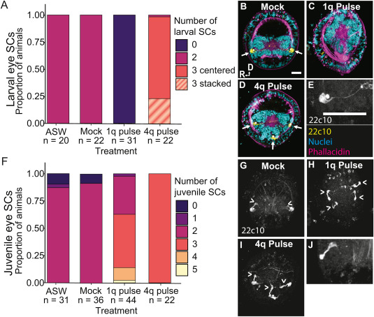
Fig. 10 Fig. 10. The effect of BMP treatment on larval and juvenile eye sensory cells (SCs). A–E) Larval eye SC phenotypes after BMP treatment. F–J) BMP increased the number of juvenile eye SCs. A) Proportion of animals with each larval eye SC phenotype in each BMP treatment. Letters indicate significance groups; colors are the same as in Fig. 9. B–E) Anterior z-stack of stage 6 larvae showing (B–D) larval SCs (22C10, yellow, arrows), nuclei (ToPRO3-iodide, teal), and muscles and microvilli (phallacidin, magenta). E) Image of single larval eye SC. F) Proportion of animals with each number of juvenile eye SCs in each BMP treatment. G–J) Juvenile eye SCs (22C10, arrowheads). B,G) Mock, C,H) 1q pulse, and E,I) 4q pulse animals. R, right side, D, dorsal, scale bars = 50 μm.
Reprinted from Developmental Biology, 478, Webster, N.B., Corbet, M., Sur, A., Meyer, N.P., Role of BMP signaling during early development of the annelid Capitella teleta, 183-204, Copyright (2021) with permission from Elsevier. Full text @ Dev. Biol.