
|
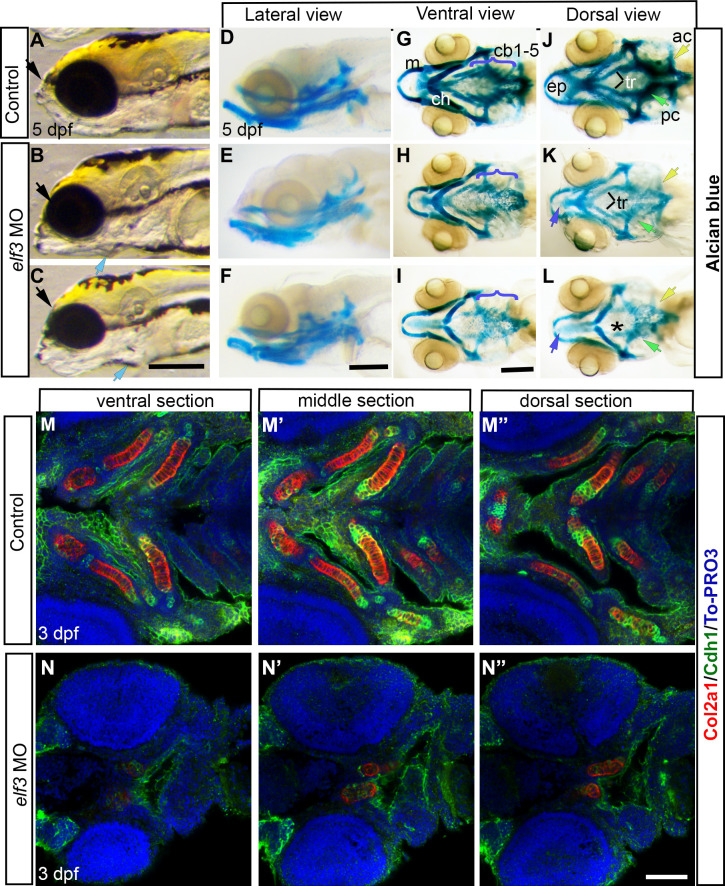
Fig 3
(A-C) Live images of 5 dpf Elf3 morphants show protruded jaw (blue arrow) and smaller head (black arrow) and eye (B, C) compared to control (A). (D-L) Alcian blue stained head skeleton of control and morphant larvae. Ventral view shows normal cartilages in control (G) and near-normal Meckel’s (m) and ceratohyal cartilages (ch) and poorly developed branchial cartilages (cb 1–5) in morphants (H, I). Dorsal view shows normal neurocranium in control (J) and smaller ethmoid plate (purple arrow), lack of trabecula (tr and *), parachordal cartilage (pc and green arrow) and auditory cartilage (ac and yellow arrow) in morphants (K, L). (M-N”) Col2α1 and E-cadherin (Cdh1) antibody stained 3 dpf larvae marked developing Meckel’s, Ceratohyal, 1st and 2nd branchial arches, and ethmoid plate in control larvae (M-M”) and a pair of cartilage (presumably Ceratohyal) in the morphant larvae (N-N”). Scale bar for A-C; D-F; and G-L = 200 μm. Scale bar for M-N” = 100 μm.