
Fig. 1
- ID
- ZDB-IMAGE-221119-35
- Publication
- Edmister et al., 2021 - A zebrafish model for calcineurin-dependent brain function
- All Figures
- Figures for Edmister et al., 2021
|
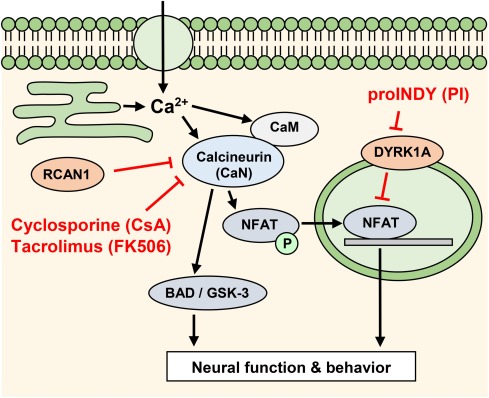
Fig. 1 Fig. 1. Model of calcineurin signaling. Intracellular free calcium activates calcineurin, which dephosphorylates various target proteins, including the nuclear factor of activated T-cells (NFAT), BCL2-associated death protein (BAD) and glycogen synthase kinase-3 (GSK-3). Calcineurin can be inhibited by the ‘Regulator of Calcineurin’, RCAN1 (previously called the ‘Down syndrome critical region’, DSCR1), or by small molecules such as cyclosporine (CsA) and tacrolimus (FK506). DYRK1A inhibits the calcineurin-NFAT pathway by phosphorylating NFAT. The small molecule INDY inhibits the inhibitor DYRK, which leads to increased NFAT signaling. ProINDY can be used as a cell-permeable prodrug to deliver INDY inside a cell. Note: NFAT, DYRK and RCAN in mammals are named nfat, dyrk, rcan (genes) or Nfat, Dyrk, Rcan (proteins) in zebrafish.