
Fig. 3
- ID
- ZDB-IMAGE-221030-9
- Publication
- Ferre-Fernández et al., 2022 - CRISPR-Cas9-mediated functional dissection of the foxc1 genomic region in zebrafish identifies critical conserved cis-regulatory elements
- All Figures
- Figures for Ferre-Fernández et al., 2022
|
Fig. 3
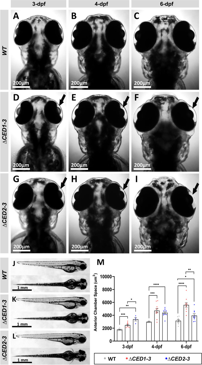
Phenotypic analysis of zebrafish mutants carrying deletions of downstream elements. A–I Dorsal images of the head region of 3-, 4- and 6-dpf wild-type (WT) (A–C), foxc1a∆CED1−3 (D–F) and foxc1a∆CED2−3 (G–I) homozygous zebrafish embryos. Both mutant lines showed the enlargement of the anterior chamber of the eye that was first noticeable at 3-dpf and became more pronounced by 6-dpf (black arrows in D–I). J–L Lateral and dorsal views of the 3-dpf wild-type (J), foxc1a∆CED1−3 (K) and foxc1a∆CED2−3 (L) homozygous zebrafish embryos. Please note no obvious morphological changes (aside from ocular defects presented in A–I) in mutant embryos. M Comparison of the anterior chamber area in wild-type and mutant embryos at 3-, 4-, and 6-dpf. *: p < 0.05; **: p < 0.01; ***: p < 0.001; ****: p < 0.0001