
Fig. 7
- ID
- ZDB-IMAGE-221030-13
- Publication
- Ferre-Fernández et al., 2022 - CRISPR-Cas9-mediated functional dissection of the foxc1 genomic region in zebrafish identifies critical conserved cis-regulatory elements
- All Figures
- Figures for Ferre-Fernández et al., 2022
|
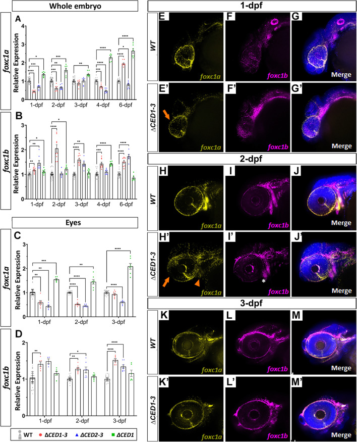
Fig. 7
Expression studies in zebrafish mutants carrying deletions of downstream regions of foxc1a. qRT-PCR relative expression of foxc1a (A, C), foxc1b (B, D) transcripts in 1–6-dpf whole bodies (A, B) and 1–3-dpf dissected eyes (C, D) of wild-type and mutant embryos; *: p < 0.05; **: p < 0.01; ***: p < 0.001; ****: p < 0.0001. E–M’ RNAscope in situ hybridization analysis of foxc1a (yellow) and foxc1b (magenta) expression in 1-, 2- and 3-dpf wild-type and foxc1a∆CED1−3 mutant embryos. Mutant embryos showed a visible reduction in ocular foxc1a expression at 1- and 2-dpf (orange arrows in E’ and H’) as well as in the branchial arches at 2-dpf (orange arrowhead in H’), while expression of foxc1b appeared normal (white asterisk; I’)