
Fig. 4
- ID
- ZDB-IMAGE-221030-10
- Publication
- Ferre-Fernández et al., 2022 - CRISPR-Cas9-mediated functional dissection of the foxc1 genomic region in zebrafish identifies critical conserved cis-regulatory elements
- All Figures
- Figures for Ferre-Fernández et al., 2022
|
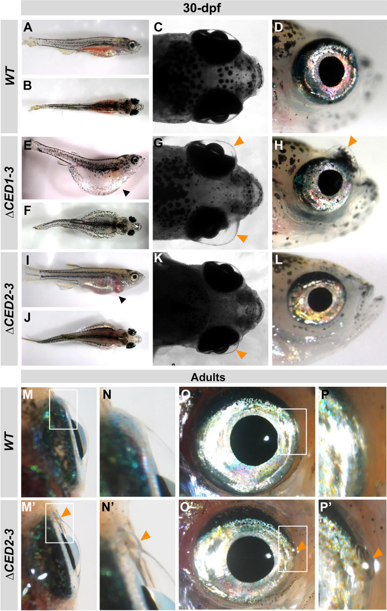
Fig. 4
Developmental defects in juvenile and adult foxc1a∆CED1−3 and foxc1a∆CED2−3 mutants. A–L Lateral and dorsal whole body and head images of 30-dpf wild-type (A–D), foxc1a∆CED1−3 (E–H) and foxc1a∆CED2−3 (I–L) homozygous zebrafish embryos. Please note general swelling, including abdominal and heart edema (black arrowheads), in mutant embryos (E, I) as well as bilateral/unilateral enlargement of the anterior chamber of the eye, particularly in the dorso-nasal region (orange arrowheads in G–H, and K). M–P’ Ocular images of adult wild-type (M–P) and foxc1a∆CED2−3 mutants (M’–P’) showing bulging in the nasal part of the anterior chamber of the eye (orange arrowheads in N’–P’). Panels N, P, N’ and P’ show the regions outlined by white boxes in panels M, O, M’ and O’ at a higher magnification