IMAGE

Fig. 2

ID
ZDB-IMAGE-221026-31
Source
Figures for Müller et al., 2022
Image
Figure Caption

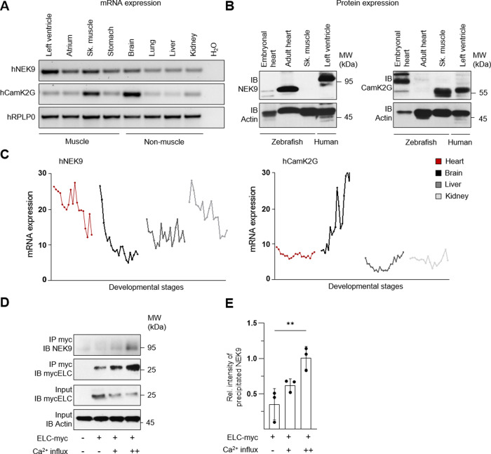
Fig. 2 Characterization of ELC interacting kinases.

A NEK9 and CamK2G RNA expression in human total RNA tissue panel. Ribosomal protein large subunit P0 (RPLP0) was used as housekeeping gene. B NEK9 (left panel) and CamK2G (right panel) protein expression in piscine and human heart tissue to detect cross-species applicability. C mRNA expression profile of NEK9 and CamK2G in different human tissues from 4 weeks post-conception to adulthood68. D, E Relationship of NEK9-ELC binding efficiency on intracellular Ca2+-concentration. NEK9-ELC interaction was analyzed after overexpression of myc-tagged ELC protein in human cells followed by specific immunoprecipitation (IP) and immunoblot (IB) analysis (D). Ca2+-influx was obtained by ionophore stimulation. Quantification of NEK9-ELC interaction dependent on Ca2+-influx illustrated as the averaged intensity of precipitated NEK9 protein normalized to actin protein expression of input protein lysate (E). Data are mean ± SD (n = 3). **P < 0.01 by the analysis of ordinary one-way ANOVA followed by Bonferroni’s multiple comparisons test.

Acknowledgments
This image is the copyrighted work of the attributed author or publisher, and ZFIN has permission only to display this image to its users. Additional permissions should be obtained from the applicable author or publisher of the image. Full text @ Nat. Commun.