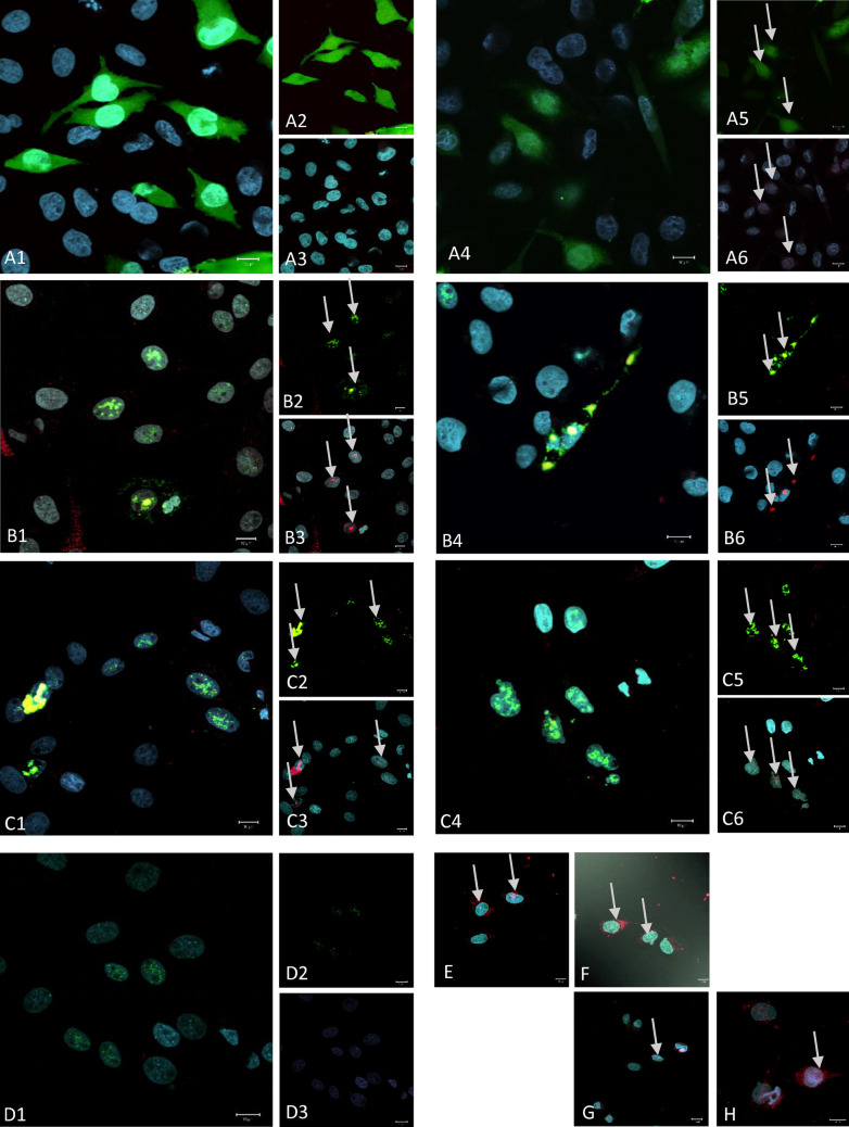
IMAGE

Figure 2

ID
ZDB-IMAGE-221018-34
Source
Figures for Klein et al., 2022
Image
Figure Caption

Figure 2

High resolution confocal imaging of single channels for the reporter fluorescence (green fluorescence proteins, green label), for immunocytochemical Bucky ball reactivity in DF1 cells after transfection with different fusion plasmids (red label) and nuclear label using SIR (cyan label): (A) two examples for the GFP only plasmid; (B) two examples for the chicken Bucky ball—venus fusion plasmid; (C) two examples for the zebrafish Bucky ball- GFP fusion plasmid; D negative control sample of chicken Bucky ball transfected cells incubated without primary Bucky ball antibody; Left larger frames represents the overlay of all three channels (1,4), right upper frames represents the overlay of the GFP reporter and Bucky ball channel (2,5), right lower frames (3,6) represent the overlay of the nuclear marker and Bucky ball label). (E,F) Examples of DF1 cells transfected with chicken Bucky ball plasmid without Venus reporter; (G,H) examples of DF1 cells transfected with zebrafish Bucky ball without GFP reporter, overlay of all three channels each; all images were taken with a 63× oil immersion 1.6 NA objective.

Acknowledgments
This image is the copyrighted work of the attributed author or publisher, and ZFIN has permission only to display this image to its users. Additional permissions should be obtained from the applicable author or publisher of the image. Full text @ Sci. Rep.