
Image
|
Figure Caption
Fig. 6
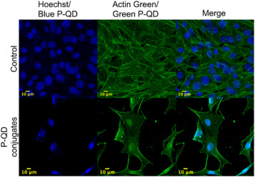
Fig. 6. Confocal microscopic images of NIH 3T3 cells. Hoechst 33342 (360/460) and Actin Green (495/518) were used as a control to stain the nucleus and cytoskeleton, respectively, and captured sequentially. Blue and green QDs were conjugated with SMAR-1 nucleus-specific protein and smooth muscle actin antibodies to target and cytoskeleton, respectively. The bottom panel was captured simultaneously at an excitation wavelength of 405 nm. Scale bar: 10 μm; Magnification: 63×.
Acknowledgments
This image is the copyrighted work of the attributed author or publisher, and
ZFIN has permission only to display this image to its users.
Additional permissions should be obtained from the applicable author or publisher of the image.
Full text @ Mater Today Bio