
Fig. 7
- ID
- ZDB-IMAGE-221001-52
- Antibodies
- Publication
- Keppeke et al., 2021 - IMPDH forms the cytoophidium in zebrafish
- All Figures
- Figures for Keppeke et al., 2021
|
Fig. 7
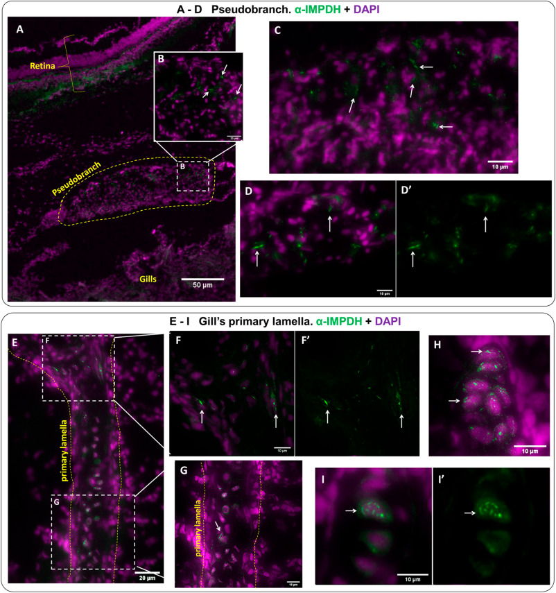
Fig. 7. IMPDH forms cytoophidia in pseudobranch and the primary lamella of the gill in adult fish. Immunofluorescence images of pseudobranch and gill of adult zebrafish cryosections for IMPDH (green) and DAPI (magenta). (A–D) Abundant cytoophidia were observed throughout the pseudobranch of the fish, highlighted in (A) by dashed lines. (B) Magnified image of selected region in (A). Arrows indicate cytoophidia in (B, C and D). (E–I) Cytoophidia were found in the primary lamella chondrocytes of the gill's cartilaginous support. In many cells cytoophidia were located in the nucleus (I). (F and G) Magnified images of selected regions in (E). Arrows indicate cytoophidia in (F–I). Scale bars = 50 μm in (A); 20 μm in (E); 10 μm in (B–D) and (F–I).
Reprinted from Developmental Biology, 478, Keppeke, G.D., Chang, C.C., Antos, C.L., Peng, M., Sung, L.Y., Coelho Andrade, L.E., Liu, J.L., IMPDH forms the cytoophidium in zebrafish, 89-101, Copyright (2021) with permission from Elsevier. Full text @ Dev. Biol.