
Fig. 3
- ID
- ZDB-IMAGE-220928-3
- Publication
- Perens et al., 2021 - osr1 couples intermediate mesoderm cell fate with temporal dynamics of vessel progenitor cell differentiation
- All Figures
- Figures for Perens et al., 2021
|
Fig. 3
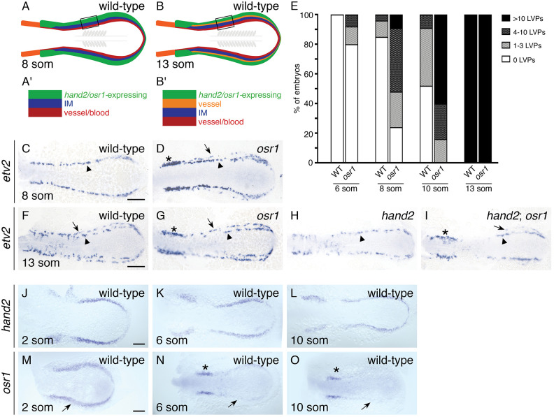
osr1 inhibits the premature emergence of lateral vessel progenitors. (A,B) Schematics depict posterior mesoderm territories, dorsal views, anterior to the left. (A′,B′) Expansion of corresponding boxed regions. (C,D,F-I) In situ hybridization shows etv2 expression in wild-type (C,F), osr1 (D,G), hand2 (H) and hand2; osr1 (I) embryos; dorsal views, anterior to the left, at the 8- (C,D) and 13- (F-I) somite stages (som). (C,D) At 8 som, etv2 is expressed in a relatively medial territory (arrowheads) on each side of the wild-type mesoderm (C). In osr1 mutants, etv2 is also expressed in some relatively lateral cells (D, arrow), and its expression is increased in a distinct proximal region (asterisk). (E) Quantification of LVPs in wild-type and osr1 mutant embryos at 6, 8, 10 and 13 som; sample sizes provided in Fig. S3E. Embryos were categorized based on the number of cells observed on whichever side of the mesoderm exhibited more LVPs. (F-I) At 13 som, etv2 is expressed in both relatively medial (arrowheads) and lateral (arrows) territories on each side of wild-type (F), osr1 (G) and hand2; osr1 (I) embryos. In hand2 mutants (H), etv2 is expressed only in the medial territory. In osr1 (G) and hand2; osr1 (I) embryos, etv2 expression is increased proximally (asterisks). (J-O) Dorsal views, anterior to the left, of in situ hybridization in wild-type embryos at 2 som (J,M), 6 som (K,N) and 10 som (L,O). Between 2 and 10 som, hand2 (J-L) remains expressed in a relatively broad domain of the wild-type posterior mesoderm. In contrast, osr1 expression is initially broad (M, arrow) but then becomes reduced in width and intensity (N,O; arrows), except proximally, where expression remains strong (asterisks), possibly representing expression in glomerular precursors (Tomar et al., 2014). Scale bars: 100 µm.