Fig. 4
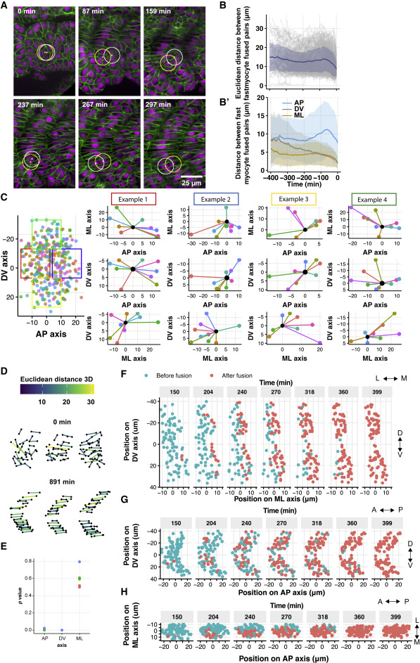
Figure 4. Fusion events cannot be predicted by initial cell position and occur in a ML wave (A) Example of fast myocyte fusion pair tracking. ML plane chosen such that the yellow highlighted cell is kept in the observed plane. The white highlighted cell is in a different ML plane in some images (hence overlap). Time t = 0 represents segmentation from the PSM. (B) Euclidean distance in 3D between fusing pairs and (B′) distance between the nucleus centroid of fusing cells along different axes. Shaded regions correspond to ±1 SD (348 cells from 3 embryos). Time t = 0 represents fusion time. (C) Left: points represent cell location immediately after segmentation for a somite. Position 0 μm in each axis corresponds to the segment center, determined by average cell position. Four regions are selected within the segment (colored boxes on left). Right: for each selected region, a cell at the same location in each of the 9 somites immediately after segmentation is identified (black point). The corresponding position of the fusing partner (other colored points) was plotted from each segment, with the position of the reference cell defined as (0,0,0). As we are comparing the local position of fusing cells, we define the selected cell to be at (0,0,0) in the (AP, DV, and ML) axes. (D) Network connections of fusing cells immediately after somite segmentation and 15 h later. Color coding represents Euclidean distance. (E) Statistical analysis (STAR Methods) of network interactions against the null hypothesis that connections are random in each axis direction. Colors represent 3 different embryos. (F–H) Nuclear position of fusing cells, color coded by their fusion state in DV-ML (F), DV-AP (G), and ML-AP (H) axes. Time defined as in (A). Position 0 μm in each axis corresponds to the segment center, determined by average cell position.
Reprinted from Developmental Cell, 57(17), Mendieta-Serrano, M.A., Dhar, S., Ng, B.H., Narayanan, R., Lee, J.J.Y., Ong, H.T., Toh, P.J.Y., Röllin, A., Roy, S., Saunders, T.E., Slow muscles guide fast myocyte fusion to ensure robust myotome formation despite the high spatiotemporal stochasticity of fusion events, 2095-2110.e5, Copyright (2022) with permission from Elsevier. Full text @ Dev. Cell

