
|
Fig. 7
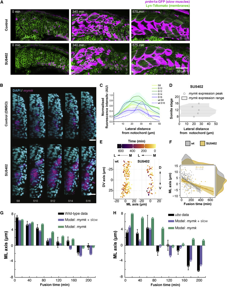
Figure 7. Spatiotemporal fusion dynamics altered by disrupting myocyte specification and rearrangements (A) Images of the developing myotome in SU5402-treated embryos during fusion, with wild-type control. Time t = 0 represents segment generation from the PSM. (B) Localization of mymk expression (magenta) visualized by FISH in an embryo treated with SU5402 (and control treated with the vehicle) and visualized at 22-somite stage co-stained with DAPI (nuclei, cyan). Scale bars, 20 μm. (C) Expression profile of mymk along the ML axis in different somite stages of SU5402-treated embryos. Data from three embryos, with intensity normalized as described in STAR Methods. Dashed lines show comparison for wild-type embryos (from Figure 5B) in S8 and S16. Position 0 μm corresponds to notochord/myotome boundary. (D) mymk expression domain (gray region, with mean ± SD shown by hexagons and error bars, respectively) from three SU5402-treated embryos in the ML axis at different somite stages. Distance as in (C). (E) Spatial distribution of fusion events in DV-ML axes in SU5402-treated embryo (right) compared with wild-type (left). Fusion events color coded by time normalized to the first fusion event per individual myotome. (F) Timing of fusion events in SU5402-treated embryos (gray) compared with wild-type (orange). The distributions show the spread of fusion timing and spatial position in ML axis (where 0 μm corresponds to the average ML position of the cells in each segment). Time t = 0 defined by the first fusion event. (E and F) 9 myotome segments from 3 embryos for wild-type and 3 myotome segments from 1 embryo in SU5402 treated. (G and H) Model predicting the timing of fusion events along the ML axis in wild-type (G) and ubo (H) embryos. Black: experimental data (from Figures 5 and 6). Purple: model prediction (STAR Methods) based on both mymk expression and slow fiber movement. Green: model prediction (STAR Methods) based on mymk expression alone. Error bars = SEM. Time and position defined as in (F).
Reprinted from Developmental Cell, 57(17), Mendieta-Serrano, M.A., Dhar, S., Ng, B.H., Narayanan, R., Lee, J.J.Y., Ong, H.T., Toh, P.J.Y., Röllin, A., Roy, S., Saunders, T.E., Slow muscles guide fast myocyte fusion to ensure robust myotome formation despite the high spatiotemporal stochasticity of fusion events, 2095-2110.e5, Copyright (2022) with permission from Elsevier. Full text @ Dev. Cell