
Image
|
Figure Caption
Fig. 5
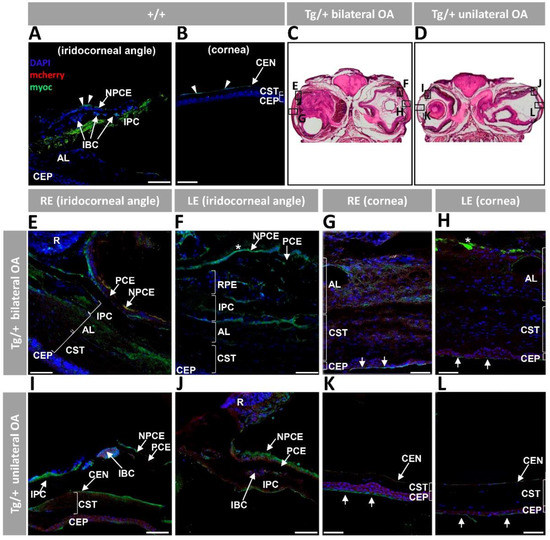
Figure 5. Fluorescent immunohistochemistry expression analysis of myocilin and mCherry in the anterior segment of old (two years) myoc transgenic zebrafish. A chicken anti-myocilin primary antibody was used to detected myocilin (green signal). Control wild type iridocorneal angle (A) and cornea (B). Representative head sectionsindicate the regions analyzed by immunohistochemistry in transgenic zebrafish with bilateral (C) or unilateral (D) ocular alterations. Anterior segment tissue sections from zebrafish with bilateral (E–H) or unilateral (I–L) ocular alterations. Asterisk in (F,H): myocilin immunoreactivity in the vitreous and iris stroma, respectively. Arrows in (G,H,K,L): myocilin immunoreactivity in the most superficial layer of the corneal epithelium. Scale bars: 50 μm; AL: annular ligament; CEN: corneal endothelium; CEP: corneal epithelium; CST: corneal stroma; IBC: iris blood cells; IPC; iris pigment cells; LE: left eye; NPCE: non-pigmented ciliary epithelium; OA: ocular alterations; PCE: pigmented ciliary epithelium; R: retina; RE: right eye; RPE: retinal pigment epithelial cells; +/+: wild type; Tg/+: transgenic.
Acknowledgments
This image is the copyrighted work of the attributed author or publisher, and
ZFIN has permission only to display this image to its users.
Additional permissions should be obtained from the applicable author or publisher of the image.
Full text @ Int. J. Mol. Sci.