
Fig. 2
- ID
- ZDB-IMAGE-220912-16
- Publication
- Zink et al., 2022 - Altered Expression of TMEM43 Causes Abnormal Cardiac Structure and Function in Zebrafish
- All Figures
- Figures for Zink et al., 2022
|
Fig. 2
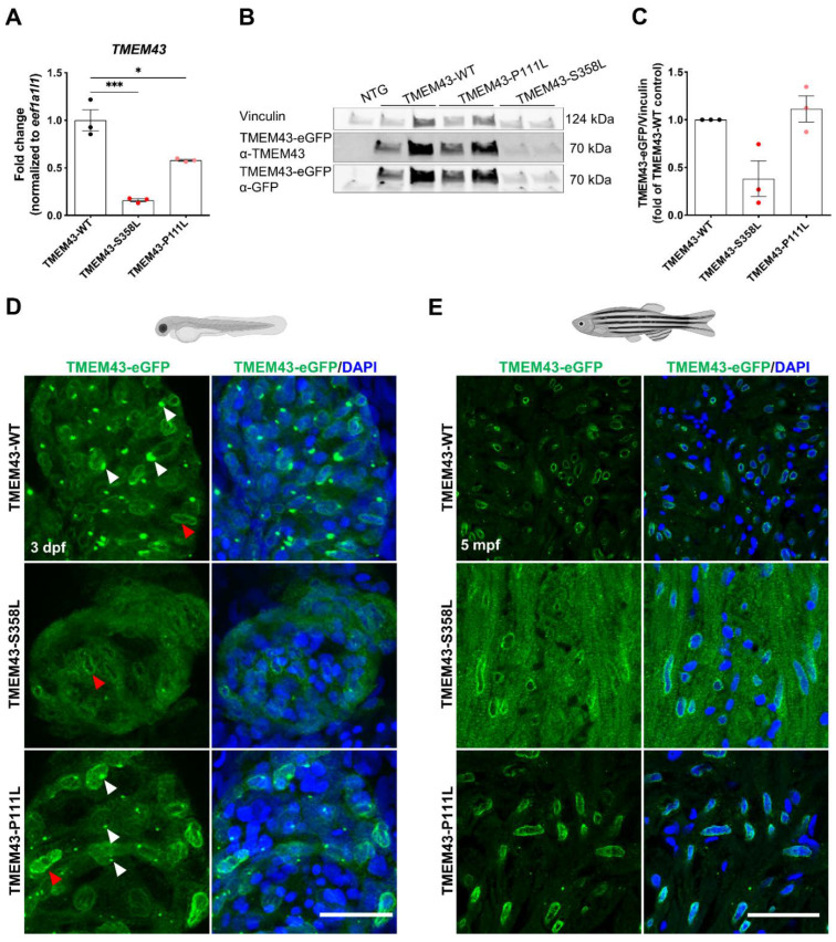
Reduced expression and altered localization of TMEM43-p.S358L. (A) Quantitative reverse transcription PCR demonstrates transcript reduction in human TMEM43 in adult ventricles of both TMEM43 variant transgenic lines compared to wild-type (WT). Each replicate consists of a pool of 2–7 ventricles and relative expression is calculated to TMEM43-WT expression levels. (B) Representative Western blot showing reduced occurrence of the human TMEM43-eGFP fusion-protein in adult ventricles of TMEM43-S358L compared to the TMEM43-WT transgenic line. For detection, both a TMEM43- and a GFP-antibody were used. Each biological replicate consists of a pool of 1–4 ventricles. (C) Western blot analysis using the TMEM43-antibody showed a reduced protein level of the human TMEM43-eGFP fusion-protein in adult ventricles of TMEM43-S358L compared to the TMEM43-WT transgenic line. Each data point represents relative Western blot band intensity of a technical replicate. (A,C) One-way ANOVA with Bonferroni’s multiple comparison test, * p ≤ 0.05, *** p ≤ 0.001. Error bars correspond to SEM. (D) Confocal images of whole-mount immunofluorescence staining of hearts expressing the transgene TMEM43-eGFP (green) at 3 days post-fertilization (dpf). Note the TMEM43-WT expression at the nuclear membrane and the endoplasmic reticulum (ER). TMEM43-S358L displays a weak signal at the nuclear membrane, but no signal at the ER. White arrowheads indicate TMEM43-eGFP localization at the ER, red arrowheads indicate localization at the nuclear membrane. Scale bar = 30 µm. (E) Confocal images of immuno-stained paraffin sections of 5 months post fertilization (mpf) dissected hearts expressing the transgene TMEM43-eGFP (green). Scale bar = 30 µm. Created with BioRender.com (accessed on 13 August 2022).