
|
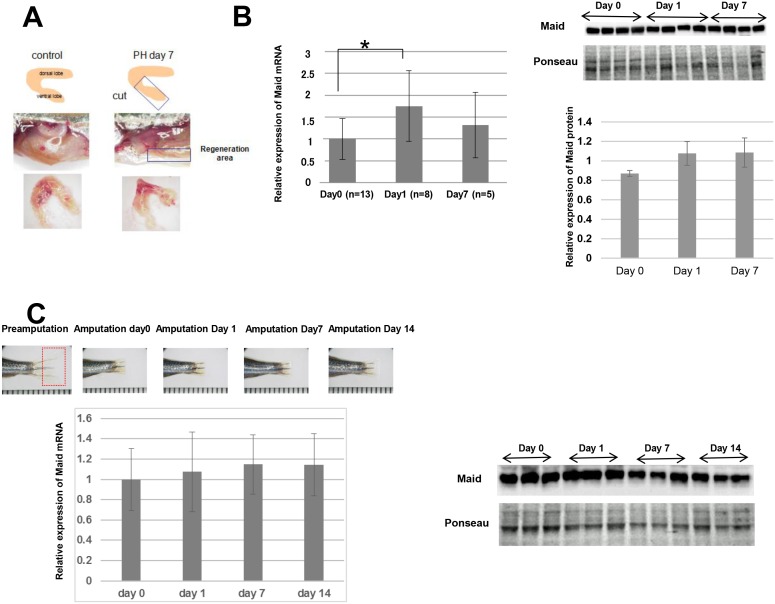
Fig 3
(A) Top: Diagram of the area of adult zebrafish liver subjected to PH. Middle: Macroscopic views of fish abdomens, sliced in the middle. About 30% of the ventral lobe of the liver was removed. The area of the liver regeneration is demarcated by a rectangle. Bottom: Macroscopic view of the livers in the middle panel after 7 days of regeneration. Results are representative of 10 fish per group. (B) Left: RT-PCR analysis of