
IMAGE
Fig. 1
- ID
- ZDB-IMAGE-220905-30
- Publication
- Moog et al., 2022 - Clemizole and Trazodone are Effective Antiseizure Treatments in a Zebrafish Model of STXBP1 Disorder
- All Figures
- Figures for Moog et al., 2022
Image
|
Figure Caption
Fig. 1
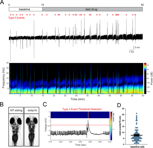
Seizure phenotype in larval stxbp1b mutant zebrafish. A, Local field potential (LFP) recording from a representative agarose-embedded stxbp1b mutant at 4 d postfertilization (dpf). Electrode is located in the optic tectum. At top is a schematic of the experimental protocol for baseline (0–15 min) and test drug exposure (15–60 min) periods. Test drug in this example is phenytoin (PHT). Type II ictal-like events are denoted by open circles (red). A 60-min continuous gap-free LFP recording trace (top) and associated spectrogram (at bottom) are shown. B, Representative images from age-matched wild-type (WT) sibling and darkly pigmented stxbp1b mutant larvae at 4 dpf. C, Example of a Type II ictal-like event identified using an amplitude threshold detector set to exclude small-amplitude brief interictal-like activity. Note the presence of a brief period of postictal depression in the electrical recording following the event. D, Plot showing the frequency of Type II ictal-like events in stxbp1b mutant larvae used for pharmacology studies (n = 84)
Figure Data
Acknowledgments
This image is the copyrighted work of the attributed author or publisher, and
ZFIN has permission only to display this image to its users.
Additional permissions should be obtained from the applicable author or publisher of the image.
Full text @ Epilepsia Open