|
Fig 4
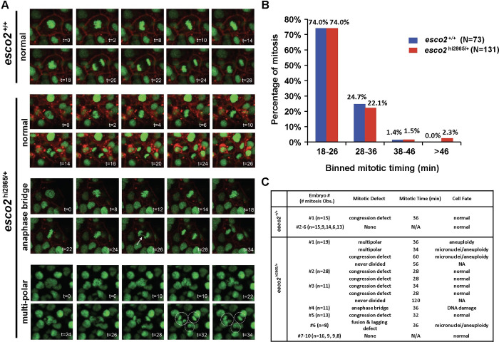
(A) in-vivo confocal imaging of H2A.F/Z-EGFP and CaaX-mCherry mRNA injected embryos at 24 hours post-fertilization (hpf) for two hours. Representative images of normal and defective mitoses in

|
|
Fig 4
(A) in-vivo confocal imaging of H2A.F/Z-EGFP and CaaX-mCherry mRNA injected embryos at 24 hours post-fertilization (hpf) for two hours. Representative images of normal and defective mitoses in