IMAGE

FIG 2

ID
ZDB-IMAGE-220903-14
Source
Figures for Deng et al., 2022
Image
Figure Caption

FIG 2

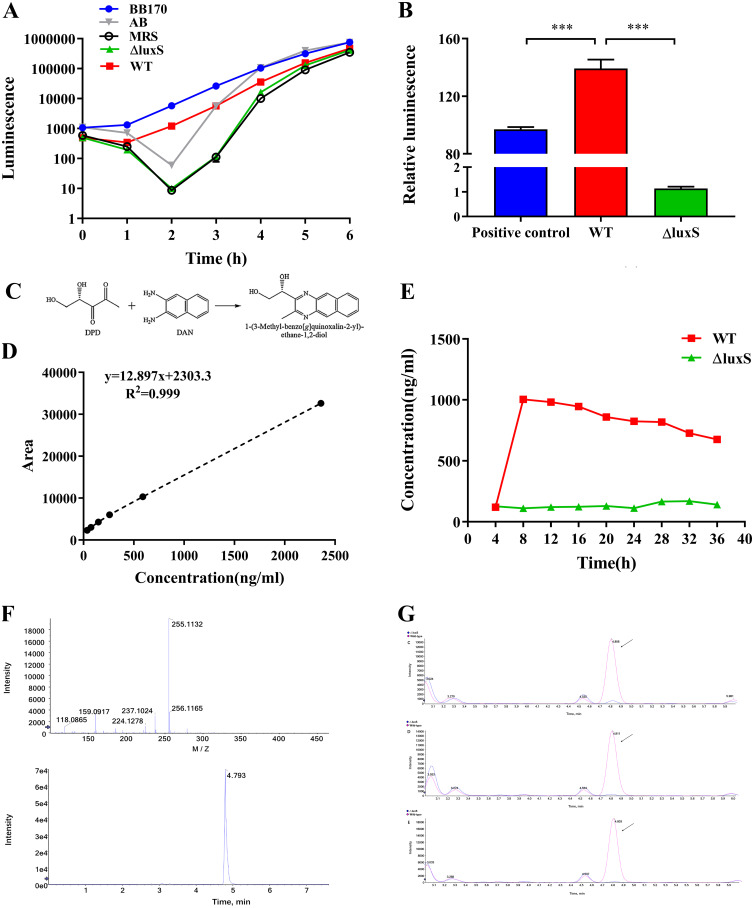
AI-2 activity of LGG wild-type or ΔluxS. (A) Detection of AI-2 activity in the supernatant of the wild-type strain and ΔluxS mutant after inducing V. harveyi BB170 to emit light. The supernatant of V. harveyi BB170, AB liquid medium, and MRS liquid medium were used as positive, negative and medium controls, respectively, n = 6. (B) Relative luminescence compared to the negative control at 24 h, n = 6. P < 0.001 (***). (C) The process of 4,5-dihydroxy 2,3-pentanedione (DPD) derivatization. It was an atypical condensation reaction with the loss of two water molecules. (D) Standard curve of AI-2 detected by HPLC-FLD. (E) Quantitative detection of AI-2 in the supernatant of LGG strains from different time points by HPLC-FLD. (F) MS spectra of the derived product of DPD. (G) Representative chromatograms of LGG supernatant (8h, 16h, 24h). WT, wild type; ΔluxS, luxS mutant; red, wild type; blue, ΔluxS strain.

Acknowledgments
This image is the copyrighted work of the attributed author or publisher, and ZFIN has permission only to display this image to its users. Additional permissions should be obtained from the applicable author or publisher of the image. Full text @ Microbiol Spectr