IMAGE

Fig. 1

ID
ZDB-IMAGE-220901-14
Source
Figures for Liu et al., 2022
Image
Figure Caption

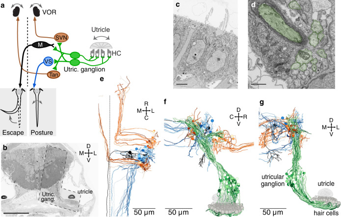
Fig. 1 High-resolution serial-section electron microscopy of the gravity-sensing system.

a Schematic of the gravity-sensing system in fish. Hair cells in the utricular macula (HC, gray) are inertial sensors of head tilt and translation, exciting the peripheral process of utricular afferents (green). These afferents, whose cell bodies are located in the utricular ganglion, project to brainstem neurons involved in escape (Mauthner cell, black), posture (vestibulospinal [VS] cell, blue), and oculomotor (VOR) reflexes (superior vestibular nucleus [SVN] and tangential nucleus [Tan], brown). Dashed line indicates midline. b Coronal section through the head of a 5.5 dpf zebrafish. The region reimaged at high resolution is visible as an L-shaped territory (dashed outline) covering the right utricle and hair cells, utricular ganglion, and ipsilateral brainstem. The reimaged territory extended across 1757 coronal sections (105 µm in the rostrocaudal axis). Scale bar, 100 µm. c Electron micrograph of two hair cells in the utricular macula, with portions of their cilia. Scale bar, 3 µm. Source data for this and all example EM images are provided as links in the Source Data file. d Section of the vestibular nerve, peripheral processes. At this developmental stage, some axons are myelinated (pseudocolored dark green) while others are not yet (light green). Scale bar, 1 µm. e Horizontal projection of reconstructed brainstem targets (Mauthner, VS, SVN, Tangential) colorized as in a. The file to generate this and all other reconstructions in this paper are provided in the Source Data file. f Sagittal projection of utricular hair cells, afferents, and brainstem targets, as in a. g Coronal projection as in f.

Acknowledgments
This image is the copyrighted work of the attributed author or publisher, and ZFIN has permission only to display this image to its users. Additional permissions should be obtained from the applicable author or publisher of the image. Full text @ Nat. Commun.