
Fig. 2
- ID
- ZDB-IMAGE-220831-24
- Publication
- Paul et al., 2022 - Aluminum (Al) causes a delayed suppression of nucleotide excision repair (NER) capacity in zebrafish (Danio rerio) embryos via disturbance of DNA lesion detection
- All Figures
- Figures for Paul et al., 2022
|
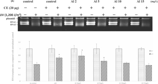
Fig. 2 Fig. 2. Downregulation of NER capacity in zebrafish embryos exposed to sublethal levels of Al. Embryos at 1 hpf were exposed to Al at the indicated concentrations for 71 h and NER capacity was detected by a transcription-based repair assay. (A) A representative gel showing the results of in vitro transcription promoted by 20 μg zebrafish extract proteins at 37 °C for 3 hr. (B) Quantitative determination of the effects of Al on NER capacity in zebrafish embryos. Each bar graph represents the mean and standard deviation of five separate experiments. *indicates p < 0.05 when compared to protein-free negative control. CE stands for crude extracts. No extract under the first two bar graphs indicates the transcriptional activities of marker cDNA in UV-irradiated and unirradiated plasmid not incubated with extract proteins.