
Image
|
Figure Caption
Fig. 3
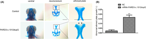
Expression of the patient-derived truncated PARD3 (c.1012dupG) variant induced hypoplastic ethmoid plate development in zebrafish. (A) Representative images of the control group and the larvae with patient-derived truncating variant mRNA injection are depicted. Compared with the development of the ethmoid plate in control group larvae, the larvae injected with PARD3-c.1012dupG mRNA showed ethmoid plate dysplasia, with the median ethmoid plate having a certain degree of absence and failing to form a smooth upper edge of the ethmoid plate. Scale bar = 200 μm. (B) Quantification of hypoplastic development of the ethmoid plate between the experimental groups and the control group (bars indicate the means ± SEM. Student’s t test was used to analyse the data. **p < 0.01)
Acknowledgments
This image is the copyrighted work of the attributed author or publisher, and
ZFIN has permission only to display this image to its users.
Additional permissions should be obtained from the applicable author or publisher of the image.
Full text @ J. Cell. Mol. Med.