
|
Fig. 11
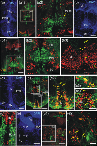
Confocal microscopic analysis of Otpa-positive cells in adult zebrafish brains of shh-GFP or islet1-GFP transgenic zebrafish line brains shows cellular co-localization of Otpa and shh-GFP in TPp-m cells (a–a2) and of Otpa and islet1-GFP in few midline pallial cells and within Vi (b-b3), massive such co-localization in Hv and some in ATN (c-c4) and Hc-a (d), as well as in PL (e-e2). Abbreviations. ATN, anterior tuberal nucleus; Hc-a, caudal zone of periventricular hypothalamus in front of posterior recess; Hv, ventral zone of periventricular hypothalamus; LH, lateral hypothalamic nucleus; llf, lateral longitudinal fascicle; NLV, nucleus lateralis valvulae; oc, optic chiasma; PL, perilemniscal nucleus; PM, magnocellular preoptic nucleus; Po, preoptic region; PPa/PPp, anterior/posterior parvocellular preoptic nucleus; PTN, posterior tuberal nucleus; PVO, paraventricular organ; RV, rhombencephalic ventricle; SC, suprachiasmatic nucleus; TPp, periventricular nucleus of posterior tuberculum; TPp-m, magnocellular cells of the periventricular posterior tubercular nucleus; Val, valvula cerebelli; Vi, intermediate nucleus of ventral telencephalon