
|
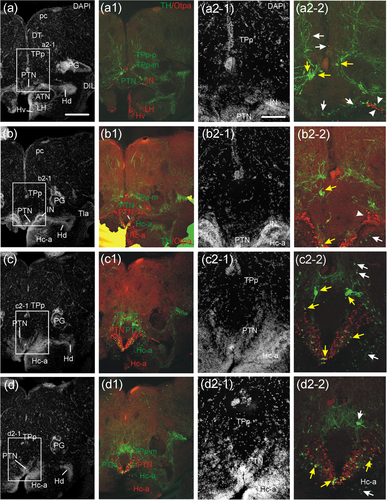
Fig. 7 Confocal microscopic analysis of catecholaminergic (TH) and Otpa-positive cells in the adult zebrafish diencephalon at levels of the posterior tuberal nucleus, intermediate hypothalamic nucleus, as well as dorsal and prerecess caudal periventricular hypothalamic zones. Four transverse levels (a-d/a2-1 to d2-1): DAPI stain; (a1-d1/a2-2 to d2-2): merged TH/Otpa immunostain) cover the rostrocaudal extent of the posterior tuberal nucleus (PTN). Note that (a2-1/a2-2) to (d2-1/d2-2) are separate enlarged microphotographs. The PTN shows rostrally TH cells (two lower white arrows in a2-2) and few Otpa-positive cells. Increasingly more Otpa-positive cells in PTN occur caudally (b2-1/b2-2 to d2-1/d2-2) where also the majority of TH/Otpa double-labeled cells in PTN sit (respective yellow arrows in b2-2 to d2-2). These three levels also show presence of TH/Otpa colabeled magnocellular cells of the periventricular posterior tuberculum (TPp-m; a2-2 to c2-2; respective yellow arrows, white arrow in [d2-2] shows a TH-only positive TPp-m cell). The parvocellular TPp-p is not double-labeled (two upper white arrows in a2-2/c2-2). The intermediate hypothalamic nucleus (IN) is characterized by a distinct Otpa-positive cell cluster, two white arrowheads in (a2-2); white arrowhead in (b2-2). Some TH-positive cells of PTN sit at the medial edge of IN, one of them double-labeled with Otpa. The caudal periventricular hypothalamic zone in front of the posterior recess (Hc-a) contains many Otpa-positive cells basal to its TH-positive cells (b1-d1). TH-positive cells in Hc-a are indicated by a white arrow in the right lower corner of (b2-2 to d2-2), note their bipolar liquor-contacting nature in (b2-2). Scale bars = 250 µm (a) applies also to (b-d) and (a1-d1), 125 µm (a2-1) applies also to (b2-1 to d2-1) and (a2-2 to d2-2). Abbreviations: ATN, anterior tuberal nucleus; DIL, diffuse nucleus of hypothalamic inferior lobe; DT, dorsal thalamus; Hc-a, caudal zone of periventricular hypothalamus in front of posterior recess; Hd/Hv, dorsal/ventral zone of periventricular hypothalamus; IN, intermediate hypothalamic nucleus; LH, lateral hypothalamic nucleus; pc, posterior commissure; PG, preglomerular complex; PTN, posterior tuberal nucleus; TLa, torus lateralis; TPp, periventricular nucleus of posterior tuberculum; TPp-m, magnocellular cells of TPp; TPp-p, parvocellular cells of TPp