
|
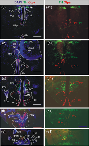
Fig. 3 Distribution of catecholaminergic (TH) and Otpa-positive cells in the adult zebrafish forebrain II. Based on DAPI stains, epifluorescence microscopic analysis of immunostained transverse sections shows no cellular colocalization of TH and Otpa in most of these catecholaminergic forebrain sites, that is, neither in the periventricular pretectum (PPr; a/a1-b/b1), the paraventricular organ (PVO; c,c1), the small-celled (parvocellular) part of the periventricular posterior tuberculum (TPp-p; c/c1), nor in the caudal zone of the periventricular hypothalamus (Hc-a/b; d/d1–e/e1). Furthermore, the ventral zone of the periventricular hypothalamus (Hv), and the anterior tuberal and lateral hypothalamic nuclei (ATN, LH) as well as the intermediate hypothalamic nucleus only contain Otpa-positive cells. However, cellular colocalization of Otpa and TH is seen in the pear-shaped magnocellular part of the posterior tuberculum (TPp-m; b/b1) and the posterior tuberal nucleus (PTN; see also Figures 7; 8). Scale bars = 250 µm. Abbreviations: A, anterior thalamic nucleus; ATN, anterior tuberal nucleus; CM, corpus mamillare; CP, central posterior thalamic nucleus; DP, dorsal posterior thalamic nucleus; Hc-a, caudal zone of periventricular hypothalamus in front of posterior recess, Hc-b, caudal zone of periventricular hypothalamus around posterior recess; Hd/Hv, dorsal/ventral zone of periventricular hypothalamus; IN, intermediate hypothalamic nucleus; LH, lateral hypothalamic nucleus; LR, lateral hypothalamic recess; PGl/PGm, lateral/medial preglomerular nucleus; PPp, posterior parvocellular preoptic nucleus; PPr, periventricular pretectum; PR, posterior hypothalamic recess; PTN, posterior tuberal nucleus; PVO, paraventricular organ; TPp, periventricular nucleus of posterior tuberculum; SCO, subcommissural organ; VL/VM, ventrolateral/ventromedial thalamic nucleus