
Image
|
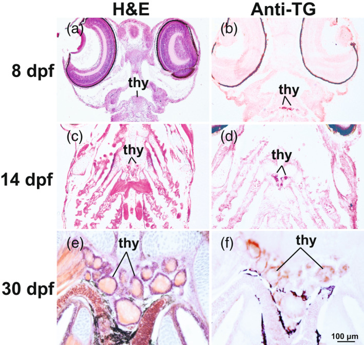
Figure Caption
Figure 3
Colocalization of thyroid follicles and thyroglobulin protein at 8, 14, and 30 dpf in stickleback larvae. (a, c, e) Hematoxylin and eosin (H&E)‐stained horizontal sections of the pharynx showing thyroid follicles at 8–30 dpf. (b, d, f) Positive staining for thyroid antibody (anti‐TG [thyroglobulin]) is visible in and around the lumen of thyroid follicles at 8–30 dpf. Imaged at 400X. thy, thyroid follicles
Acknowledgments
This image is the copyrighted work of the attributed author or publisher, and
ZFIN has permission only to display this image to its users.
Additional permissions should be obtained from the applicable author or publisher of the image.
Full text @ Evol. Appl.