
Fig. 3
- ID
- ZDB-IMAGE-220720-40
- Publication
- Lin et al., 2022 - Macrophages Break Interneuromast Cell Quiescence by Intervening in the Inhibition of Schwann Cells in the Zebrafish Lateral Line
- All Figures
- Figures for Lin et al., 2022
|
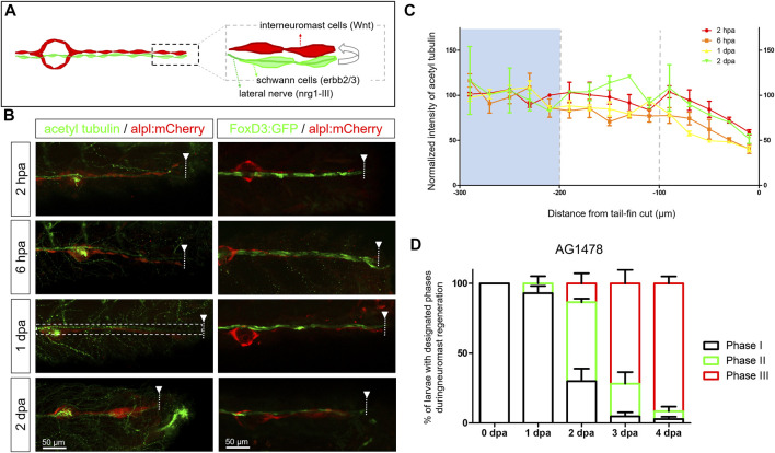
Fig. 3
Diminished nerve inhibition near the cut site may allow neuromast regeneration from interneuromast cells. (A) A graph shows the close contact between interneuromast cells (INCs, red) with underlying Schwann cells (SWCs, light green) and posterior-lateral nerve (pLLn, dark green). The pLLn-derived nrg1-III can activate erbb2/3 receptors on SWCs to suppress Wnt activity in INCs, thus keeping INCs in a quiescent state. (B) The 3-dpf larvae from the cross of Tg(−4.7alpl:mCherry) and Tg(foxd3:EGFP) were tail fin–amputated and immobilized at the designated time to show the integrity of SWCs in green [labeled by Tg(FoxD3:GFP) in all stages examined (right column)]. Tg(−4.7alpl:mCherry) larvae at 3 dpf were also tail fin–amputated, fixed at designated hours or days post-amputation (hpa or dpa, respectively), and subjected to immunostaining against acetyl tubulin to reveal green nerves along the red lateral line (left column). The fluorescent intensity of green pLLn is diminishing, approaching the cut site (dashed line). So, we measured the green fluorescent intensity at decreasing distance from the cut site in cropped images as depicted by a dotted rectangle as shown in the image of a 1-dpa larva. The averaged intensity was calculated and normalized with those of the proximal region (200–300 μm from cutting edge, blue background) for designated times shown by different colors. (D) The Et(HG7L) larvae at 3 dpf were treated with 3 μM of AG1478 and fin-amputated to examine neuromast regeneration according to Figure 1C. Data are presented as mean ± s.e.m.