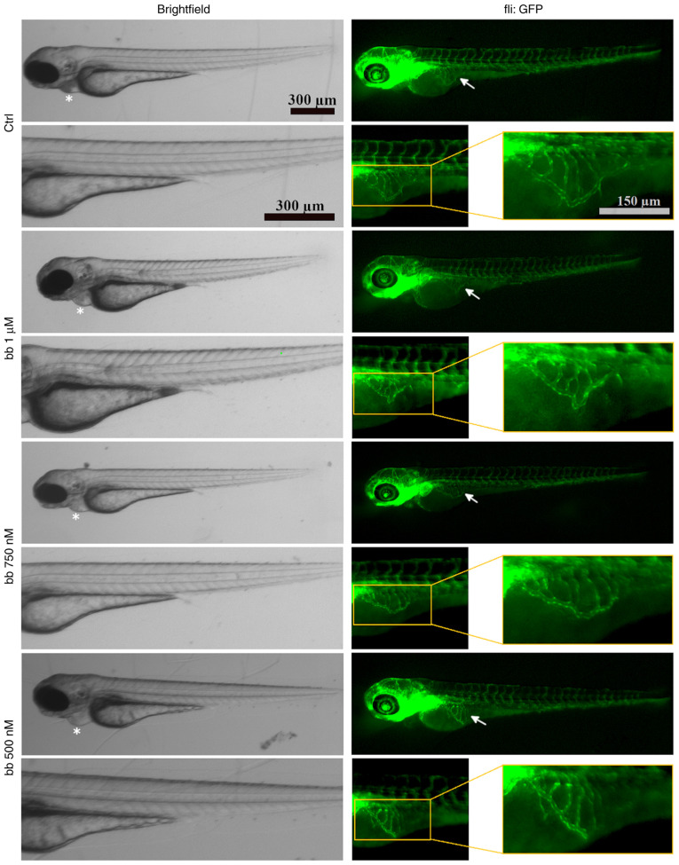
Fig. 11
Morphology of the blood vessel system of transgenic Tg(fli1:EGFP)y1 mutant Casper zebrafish embryos 72 h post-fertilisation. Embryos were treated for 48 h with either a corresponding amount of DMSO, with bb concentrations of 1 µM, 750 or 500 nM. Brightfield and green fluorescent images, denoted as fli:GFP, are shown. The pericardial region was marked with asterisks whereas SIVs are marked with arrows. Fluorescent SIV areas are marked with yellow boxes, which were then magnified two-fold magnification. Individuals shown are representative for ≥20 embryos per concentration. bb, broxbam; GFP, green fluorescent protein; SIV, sub-intestinal veins.

