IMAGE

Figure 3

ID
ZDB-IMAGE-220709-22
Source
Figures for Xu et al., 2022
Image
Figure Caption

Figure 3

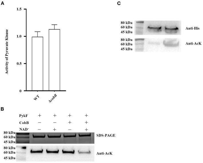
(A) CobB deacetylation PykF in vitro. Top line, Western blot was performed on PykF treated with CobB for 1 h. The same amount of protein was loaded in all lanes. Bottom line, pyruvate kinase activity was measured, and the enzyme activity of lane 1 sample was set as 1. Mean values and standard deviations were calculated based on three replicates (n = 3). Two-tailed P-values were determined by the t-test, and the significance level is 0.05. (B) CobB deacetylates PykF in vivo. Top line, Western blot analysis of purified PykF from V. alginolyticus WT:pykF and ΔcobB:pykF. Lane 1, purified PykF from V. alginolyticus WT:pykF; Lane 2, purified PykF from V. alginolyticus ΔcobB:pykF. The same amount of protein was loaded in all lanes. Anti-His, His Tag Mouse Monoclonal Antibody. Anti-AcK: anti-acetyl lysine antibody. Bottom line, pyruvate kinase activity of V. alginolyticus WT and ΔcobB. Pyruvate kinase activity of V. alginolyticus WT was set as 1. Mean values and standard deviations were calculated based on three replicates (n = 3). Two-tailed P-values were determined by the t-test, and the significance level is 0.05.

Acknowledgments
This image is the copyrighted work of the attributed author or publisher, and ZFIN has permission only to display this image to its users. Additional permissions should be obtained from the applicable author or publisher of the image. Full text @ Front Vet Sci