
|
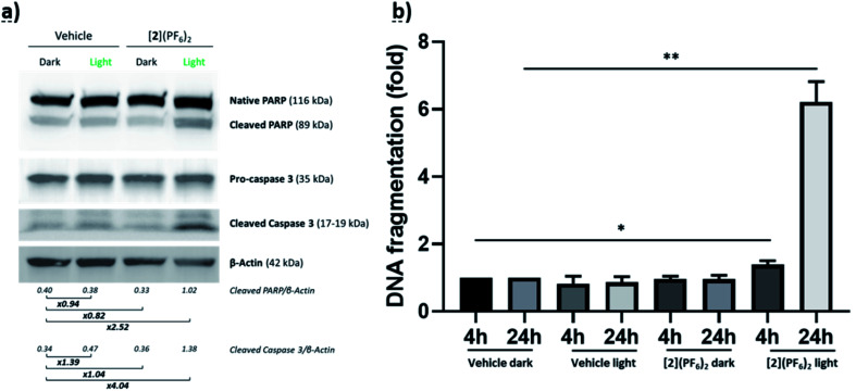
Fig. 6 Apoptosis-associated proteins and DNA fragmentation in CRMM1 cells treated with [2](PF6)2. (a) Western blot showing native PARP, cleaved PARP, pro-caspase-3, cleaved caspase-3 and β-actin as loading control in CRMM1 cells treated with [2](PF6)2 at EC50,light (4 μM) for 24 h, irradiated or not with green light (520 nm, 15 min, 21 mW cm−2, 19 J cm−2), and further incubated for 48 h in the dark. (b) DNA fragmentation in CRMM1 cells treated with [2](PF6)2 at EC50,light (4 μM) for 24 h, irradiated or not with green light (520 nm, 15 min, 21 mW cm−2, 19 J cm−2), and incubated in the dark for 4 h or 24 h after light irradiation.