
|
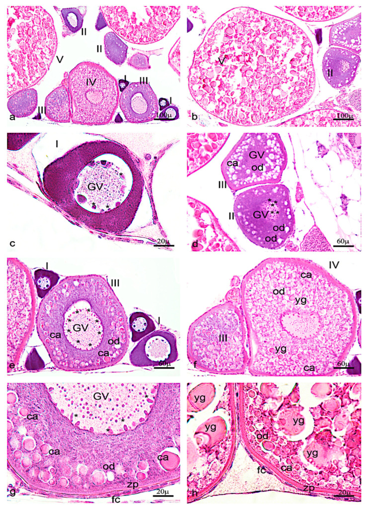
Fig. 6
Hematoxylin-eosin staining of adult zebrafish ovary. (a) Overview of adult zebrafish ovary by hematoxylin-eosin staining, five oocyte stages (I-II-II-IV-V). (b) Oocyte stages II and V. (c) Oogonia characterized by large euchromatic germinal vescicle (GV), and several nucleoli peripherically located. (d) Oocyte during primary growth (stages II–III), characterized by an increase of nucleoli in GV, and in ooplasma were present oil droplets (od) around the GV. (e) Oocyte (stages III) characterized by numerous nucleoli at the periphery of GV. (f) numerous oil droplets and cortical alveoli (ca). (g) The oocyte (stages IV) is enveloped by zona pellucida (zp) and a single layer of follicular cells (fc). (h) Oocyte (stage V) characterized by a significant increase of number and size of the yolk globules (yg). Significant increase in the thickness of the zona pellucida and appearance of thecal layer. Scale bars are: 100 µ (a,b); 60 µ (d–f); 20 µ (c,g,h).