
Image
|
Figure Caption
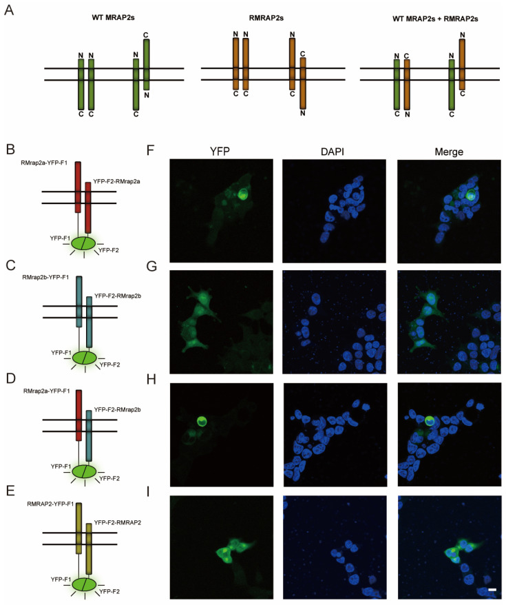
Fig. 3
RMrap2a/b and RMRAP2 form antiparallel dimers on the plasma membrane. (A) Schematic illustration of parallel and antiparallel dimers of WT MRAP2s (left), RMRAP2s (middle) as well as WT MRAP2 and RMRAP2 dimers (right). (B–E) The schematic diagrams illustrate the principle of YFP fluorescence emission and the localization of YFP-F1/F2 on the fused protein. Red: RMrap2a, blue: RMrap2b, yellow: RMRAP2. (F–I) YFP fluorescent and DAPI under confocal microscope. Scale bar = 10 μm.
Acknowledgments
This image is the copyrighted work of the attributed author or publisher, and
ZFIN has permission only to display this image to its users.
Additional permissions should be obtained from the applicable author or publisher of the image.
Full text @ Biology (Basel)