
|
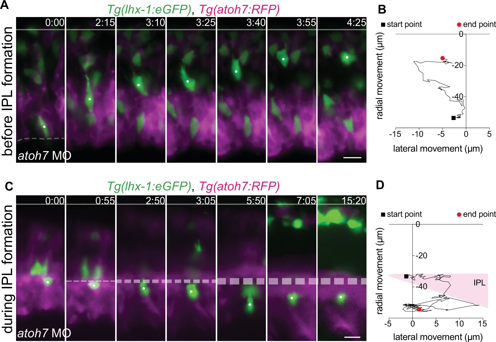
Fig. 6 - supplement 1
(A–D) Representative time series of tracked horizontal cells (HCs) (white dot) and their respective trajectories in atoh7 MO retinae at different developmental stages: before inner plexiform layer (IPL) formation (A–B), and after maturation of IPL (C–D). Tg(lhx-1:eGFP) labels HCs (green), Tg(atoh7:RFP) labels membranes of photoreceptors (PRs) and basal neurons (magenta). (A) Before IPL formation, HCs successfully move toward the HC layer even when moving from more basal positions close to the lens. (B) Radial-tangential migration trajectories of the tracked HC from (A). See Figure 6—source data 1. (C) After IPL formation, HCs fail to move through the formed IPL and therefore remain ectopically trapped beneath the IPL. (D) Radial-tangential migration trajectories of the tracked HC from (C). See Figure 6—source data 1. Line: apical surface; dashed line: forming IPL (B). Scale bar: 10 μm. Time in h:min. (A,C).
Analysis of role of IPL formation on HC migration.