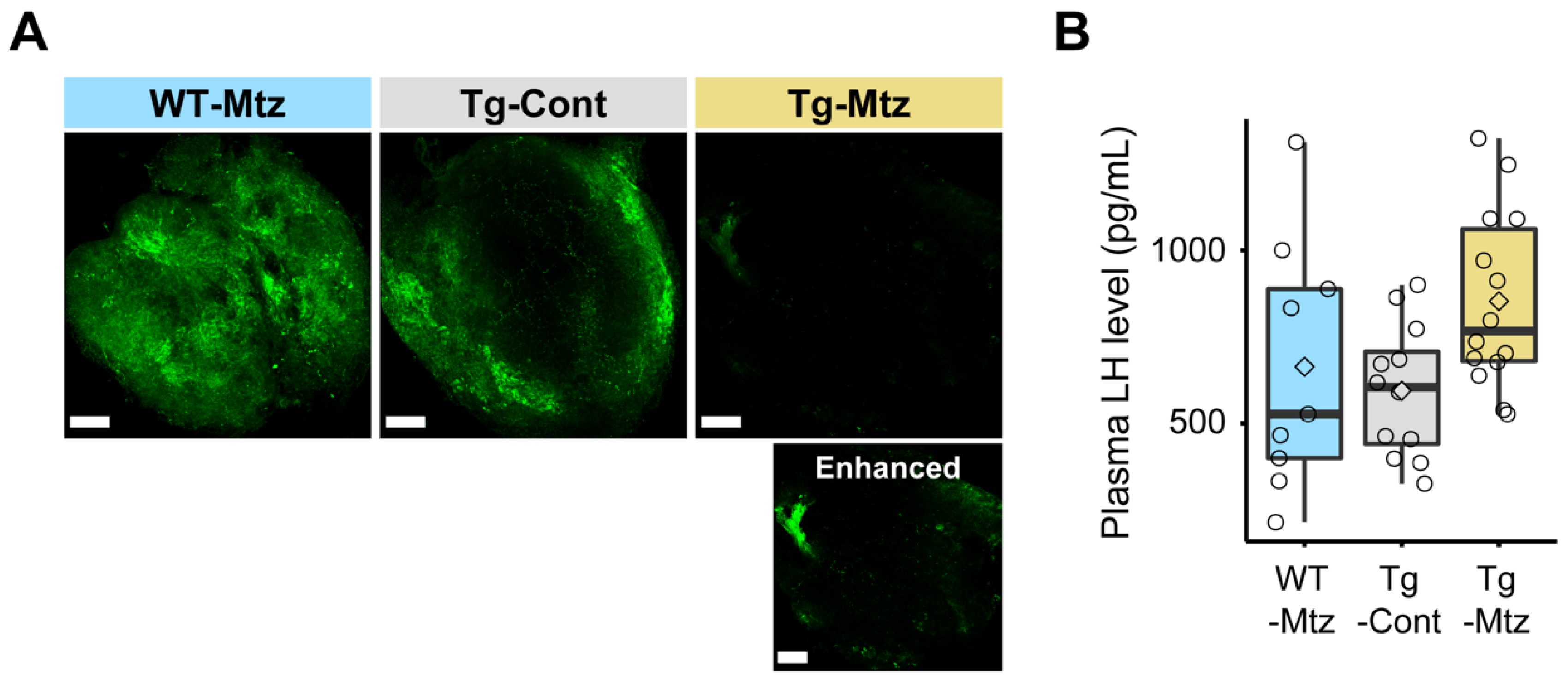
GnRH3 neuronal projections are reduced at 28 days of treatment in the pituitary of GnRH3 neuron-ablated females. (A) Confocal images of immunostaining with GnRH3-associated peptide antibody in the whole pituitary (posterior to top left), showing GnRH3 neuronal projections (green). Scale bars, 1 mm. The bottom panel of Tg-Mtz, an image with enhanced signals with levels and tone curve adjustments to demonstrate that GnRH3 is still detectable in the pituitary. (B) LH levels in plasma (n = 9–14). WT-Mtz, WT siblings treated with 2 mM metronidazole (Mtz); Tg-Cont, Tg(gnrh3:Gal4ff; UAS:nfsb-mCherry) treated with vehicle; Tg-Mtz, Tg(gnrh3:Gal4ff; UAS:nfsb-mCherry) treated with 2 mM Mtz. p > 0.05.
Acknowledgments
This image is the copyrighted work of the attributed author or publisher, and
ZFIN has permission only to display this image to its users.
Additional permissions should be obtained from the applicable author or publisher of the image.
Full text @ Int. J. Mol. Sci.
Your Input Welcome
Thank you for submitting comments. Your input has been emailed to ZFIN curators who may contact you if
additional information is required.
Oops. Something went wrong. Please try again later.