
Fig. 7
- ID
- ZDB-IMAGE-220520-24
- Publication
- Pan et al., 2022 - Gnetum montanum extract induces apoptosis by inhibiting the activation of AKT in SW480 human colon cancer cells
- All Figures
- Figures for Pan et al., 2022
|
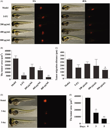
Fig. 7 Figure 7. Effect of GME on xenograft zebrafish tumour model. (A) The SW480 cells were microinjected into zebrafish embryos (larvae stage, n = 10 per group). Fluorescence area was captured by fluorescence microscopy at 0 hpf and 48 hpf after treated with GME and 5-FU. (B) Compared with the model group, the tumour area was reduced in the treatment group. *p < 0.05; **p < 0.01. (C) Compared with the model group, the tumour migration distance was reduced in the treatment group. *p < 0.05. (D) Fluorescence area of the xenograft zebrafish tumour treated by GME (400 μg/mL) for 3 dpf and 5 dpf. (E) The tumour area significantly reduced, showing in a time-dependent manner. *p < 0.05; **p < 0.01.