
Image
|
Figure Caption
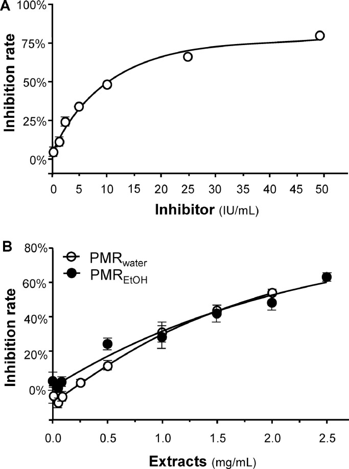
Fig. 2
Inhibition of S-protein-hACE2 binding. S-protein-hACE2 binding was analysed by ELISA. (A) Line graph showing the response of a standard inhibitor (calibrated to NIBSC code 20/136), which was used as a positive control. (B) S-protein-hACE2 binding was inhibited by PMRwater and PMREtOH in a dose-dependent manner. The inhibition percentage was determined from the binding signal normalized to the interaction between spike RBD and hACE2 without extract. The data represent mean ± SD (
Acknowledgments
This image is the copyrighted work of the attributed author or publisher, and
ZFIN has permission only to display this image to its users.
Additional permissions should be obtained from the applicable author or publisher of the image.
Reprinted from Phytomedicine : international journal of phytotherapy and phytopharmacology, 102, Wang, X., Lin, S., Tang, R.W., Lee, H.C., Chan, H.H., Choi, S.S.A., Leung, K.W., Webb, S.E., Miller, A.L., Tsim, K.W., Polygoni multiflori radix extracts inhibit SARS-CoV-2 pseudovirus entry in HEK293T cells and zebrafish larvae, 154154, Copyright (2022) with permission from Elsevier. Full text @ Phytomedicine