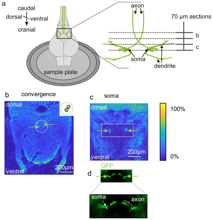
Spatial localization of a central command neuron within tissue sections of zebrafish using MALDI MSI. (a) Schematic representation of a zebrafish brain fixated on a sample holder, with the central command neurons (the pair of Mauthner neurons) shown in green. On the right side the two Mauthner neurons are magnified and the position of two sections (70 µm thickness) are illustrated with dotted lines. (b,c) Positive ion MS images. (b) MS image of PC (36:1) [M+H]+ visualizing the axon arrangement before the point where the axons cross the midline. Axons are labeled with green arrows and a schematic illustration of the axon region is shown on the right side of the MS image. (c) MS image of the somata of the two neurons (labeled with green arrows), visualized by the distribution of PC (38:6) [M+H]+. (d) Fluoresence microscopy of tissue section from transgenic line Tol056 (that expresses GFP in the neuron) was used to conveniently select appropriate sections for subsequent MSI.
Acknowledgments
This image is the copyrighted work of the attributed author or publisher, and
ZFIN has permission only to display this image to its users.
Additional permissions should be obtained from the applicable author or publisher of the image.
Full text @ Sci. Rep.
Your Input Welcome
Thank you for submitting comments. Your input has been emailed to ZFIN curators who may contact you if
additional information is required.
Oops. Something went wrong. Please try again later.