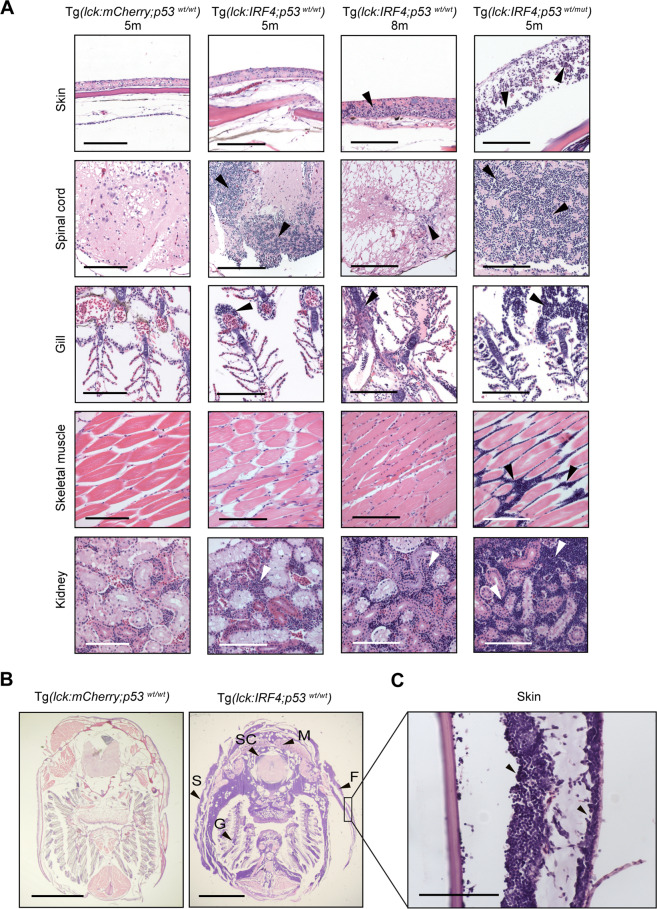
IRF4-driven zebrafish tumors recapitulate invasive human T-cell lymphoma.
A Histopathological examination of H&E-stained tissue sections of representative samples from control Tg(lck:mCherry;p53wt/wt) (n = 8), Tg(lck:IRF4;p53wt/wt) (n = 8) and Tg(lck:IRF4;p53wt/mut) (n = 4) zebrafish at 5 or 8 months. Similar findings were observed in multiple independent animals as shown for each sample. Tumor cells are indicated by white or black arrowheads. Scale bar = 100 μm. B Transverse sections of representative samples from Tg(lck:mCherry;p53wt/wt) (n = 8) and Tg(lck:IRF4;p53wt/wt) (n = 8). F, fin; G, gill; M, muscle; S, skin; and SC, spinal cord. Similar findings were observed in multiple independent animals as shown for each sample. Scale bar = 2 mm. C High magnification of skin lesions from Tg(lck:IRF4;p53wt/wt) fish. Scale bar = 100 μm.
This image is the copyrighted work of the attributed author or publisher, and
ZFIN has permission only to display this image to its users.
Additional permissions should be obtained from the applicable author or publisher of the image.
Full text @ Nat. Commun.
Your Input Welcome
Thank you for submitting comments. Your input has been emailed to ZFIN curators who may contact you if
additional information is required.
Oops. Something went wrong. Please try again later.