IMAGE

Fig. 3

ID
ZDB-IMAGE-220505-48
Source
Figures for Gabler et al., 2022
Image
Figure Caption

Fig. 3

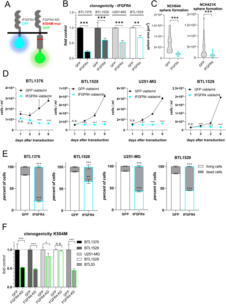
Inactivation of FGFR4 reduces proliferative capacity and promotes cell death. A Schemes of the kinase domain-truncated, CFP-coupled tFGFR4 (left) and of the point-mutated (mut), kinase-dead FGFR4-KD(K504M) (right), GFP-coupled proteins are shown. (B + F) Clonogenicity upon tFGFR4B, FGFR4-KD(K504M)(F) compared to GFP(B + F, set to 1) transduction was analyzed in endogenously FGFR4high (BTL1376, BTL1528) or FGFR4low (U251-MG, BTL1529, BTL53) cell models (mean ± SD from three experiments). C Sphere formation potential of GSC models upon tFGFR4 or GFP transduction (mean ± 25%CI). D Survival/proliferation capacities over time are shown for tFGFR4- and GFP-transduced GBM models. For each model, one representative out of three experiments in duplicates is depicted. E Cell death induction upon tFGFR4 or GFP transduction in GBM models after 3 days. Annexin/PI staining was analyzed by flow cytometry. Significance levels were calculated comparing tFGFR4- to GFP-transduced cells on the respective living or dead fraction. Statistical analyses: 2-way ANOVA/Bonferroni correction (mean ± SD) (B,D,E,F); Student’s t-tests (C). *p < 0.05, **p < 0.01, ***p < 0.001, n.s. = not significant

Acknowledgments
This image is the copyrighted work of the attributed author or publisher, and ZFIN has permission only to display this image to its users. Additional permissions should be obtained from the applicable author or publisher of the image. Full text @ Acta Neuropathol Commun