IMAGE

Figure 5

ID
ZDB-IMAGE-220430-41
Source
Figures for Li et al., 2022
Image
Figure Caption

Figure 5

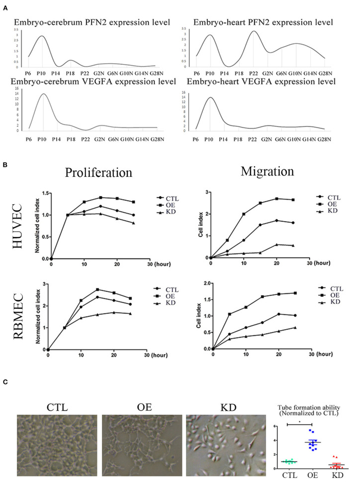
PFN2 promotes proliferation and migration of ECs, as well as tube formation and reaches the maximum level in 10-day embryo. (A) The mRNA levels of PFN2 and VEGFA at different developmental stages of gerbils (n = 15) embryo (prenatal), brain and heart (postnatal). (B) PFN2 significantly promoted proliferation of HUVECs (n = 3) and RBMECs (n = 3), as well as showed a positive effect on migration of HUVECs (n = 3) and RBMECs (n = 3). (C) The tube length in PFN2-overexpressing ECs (OE) was significantly increased than that in blank CTL and PFN2 knockdown ECs (KD), n = 3. *p < 0.05, one-way ANOVA and repeated-measures ANOVA (Tukey).

Acknowledgments
This image is the copyrighted work of the attributed author or publisher, and ZFIN has permission only to display this image to its users. Additional permissions should be obtained from the applicable author or publisher of the image. Full text @ Front Cardiovasc Med