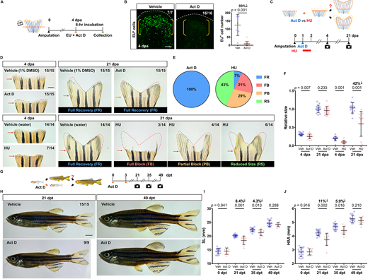
Fig. 6 Pharmacological inhibition of bulk RNA transcription reversibly pauses regeneration and growth in zebrafish. (A) Timeline of actinomycin D (Act D) treatment and EU assay during tailfin regeneration. Orange arrowhead marks the EU and Act D injection time. Green arrowhead marks the collection time. (B) Whole-mount EU staining of the wild-type tailfin regenerates at 4 dpa after a 6-h treatment of Act D. White dashed lines outline the distal margin of the fin tissue. Yellow brackets mark the blastema compartment. Scale bar: 50 μm. Graph shows quantification of EU+ cells in the lateral 2nd bony ray (n=7 vehicle, 16 Act D; mean±s.d.; Mann–Whitney U test). (C) Timeline of Act D and HU treatment during tailfin regeneration. In the Act D group, individuals at 1 dpa were intraperitoneally injected with either vehicle or Act D, as indicated by a blue arrowhead. In the HU group, individuals at 1 dpa were incubated in either vehicle or HU for 24 h, as indicated by a red bar. Fin regenerates were imaged at 4 and 21 dpa. (D) Whole-mount images of the Act D/HU-treated WT fin regenerates at 4 and 21 dpa. Red arrows indicate amputation plane. Red dashed lines depict respective fin region prior to the amputation to facilitate visual comparison. Scale bar: 1 mm. (E) Pie charts summarizing proportions of fin regenerate phenotypes at 21 dpa. Left: All of the regenerates in the Act D group (15/15) fully recovered. Right: Fin regenerates in the HU group displayed high variation. Only 7% of the animals (1/14) fully recovered. FB, full block; FR, full recovery; PB, partial block; RS, reduced size. (F) Quantification of the relative fin regenerate size. Data from each individual were normalized to their original fin sizes (n=15 vehicle, 15 Act D; 14 vehicle, 14 HU; mean±s.d.; two-tailed Student's t-test). (G) Timeline of Act D treatment in juvenile zebrafish. Individuals at 11 weeks of age were intraperitoneally injected twice with either vehicle or Act D, as indicated by orange arrowheads. Images were captured at 21, 35 and 49 days after the first treatment (dpt). (H) Whole-mount images of the vehicle- and Act D-treated zebrafish at 21 and 49 dpt (stitched). Scale bar: 2 mm. (I,J) Measurements of SL and HAA at 0, 21, 35 and 49 dpt (n=15 vehicle, 9 Act D; mean±s.d.; two-tailed Student's t-test).
Image
Figure Caption
Figure Data
Acknowledgments
This image is the copyrighted work of the attributed author or publisher, and
ZFIN has permission only to display this image to its users.
Additional permissions should be obtained from the applicable author or publisher of the image.
Full text @ Development

