
Figure 3
- ID
- ZDB-IMAGE-220426-75
- Publication
- Lane et al., 2021 - Steroid-sensitive nephrotic syndrome candidate gene CLVS1 regulates podocyte oxidative stress and endocytosis
- All Figures
- Figures for Lane et al., 2021
|
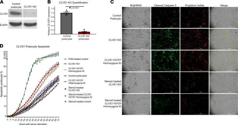
Figure 3
(A and B) Western blot showing significantly reduced expression of CLVS1 in CRISPR/Cas9-mediated knockout (KO) podocyte cell lines compared with control cell lines (n = 4, P < 0.001, 2-tailed t test). (C) Still images depicting cleaved caspase-3 activity (green) as reporter for early apoptosis and propidium iodide staining (red) for late apoptosis and necrosis in human podocytes 72 hours after serum starvation (scale bars = 1 mm). (D) Quantification of these images over time revealed an increase in podocyte susceptibility to serum starvation–induced apoptosis in CLVS1-KO and homozygous H310Y-knockin (KI) podocytes compared with controls (P < 0.05 for all time points after 16 hours for KO and after 34 hours for KI, n > 25 for each group, 2-way ANOVA). Heterozygous H310Y-KI podocytes displayed similar apoptotic phenotypes to controls. The elevated apoptosis in CLVS1-KO and homozygous H310Y-KI podocytes was rescued by treatment with 1 μM dexamethasone.