IMAGE

Figure 6

ID
ZDB-IMAGE-220416-9
Source
Figures for Garg et al., 2022
Image
Figure Caption

Figure 6

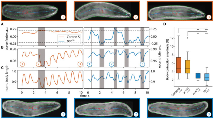
Quantification of body peristaltic contractions of freely crawling Drosophila larvae. The results of two trajectories traced with LACE are shown in (A–C): (A) the curvature, (B) eccentricity, and (C) normalized body length of a wild-type (CantonS) larva (orange) and a nan36a mutant larva (blue). The curve finder (A) detects portions of the video where turning is detected. The turns appear as gray shaded areas (point 2 for CantonS and points 2 and 3 for nan36a). The white background shows peristaltic contractions during forward crawling (points 1 and 3 for CantonS and 1 for nan36a). Above (wildtype) and below (nan36a) still frames from the corresponding times (1,2,3) are depicted. The pseudo-skeleton is superimposed as a light blue line, the contour of the animal is shown as solid green line, the central contour as a dashed green line, and the gut as a red line. Both markers (gut, central contour) were not used in this analysis. In (D) the contraction amplitude is quantified for wildtype, w1118 , nan36a and iav1 mutant larvae. The nan36a and iav1 mutants have significantly lower body contraction amplitudes compared to wildtype CantonS and w1118. The dataset consists of 30 wildtype larvae (CantonS), 26 w1118 larvae, 8 nan36a larvae, and 12 iav1 larvae. Statistical significance was tested with Fisher's permutation test on different medians. ***p < 0.001, **p < 0.01.

Acknowledgments
This image is the copyrighted work of the attributed author or publisher, and ZFIN has permission only to display this image to its users. Additional permissions should be obtained from the applicable author or publisher of the image. Full text @ Front. Behav. Neurosci.