
|
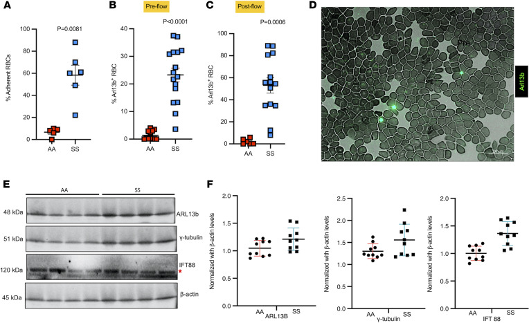
Figure 4
Sickle RBCs adhere to brain ECs triggering deciliation, and cilia are found on sickle RBCs and plasma from SCD. HBMVECs exposed to sickle (SS) or healthy (AA) RBCs were subjected to shear stress (1 dyne/cm2), and fraction of SS (n = 6) and AA (n = 5) RBCs adhered to ECs after stress induction was calculated, P = 0.0081 (A). SS and AA RBCs were tested for ARL13b cilia prior to flow and proportion of ARL13b cilia adhered to circulating SS RBCs (n = 16) versus AA RBCs (n = 12) were quantified, P < 0.0001 (B). After flow, ARL13b expression on SS RBCs (n = 11), but not AA RBCs (n = 6), upon interaction with ECs, P = 0.0006 (C). (A–C) Mann-Whitney-Wilcoxon test P values are provided. Representative field (magnification 63×; scale bar = 20 μm) of a smear of SS RBCs shows cilia presence on these sickle cells, detected with FITC-conjugated anti-Arl13b antibody (D). Western blot plot shows the detection of cilia-specific proteins in plasma samples of healthy controls (AA) versus sickle (SS). Red asterisk represents the top IFT88 band that was used for quantification (E). Please note that Western blots from only 4 AA and SS samples are shown in E. A separate gel for the other 6 samples was run and quantified. Quantification includes all 10 samples from each group. Cilia-specific proteins were quantified from plasma samples of healthy controls (AA) (n = 10) versus sickle (SS) (n = 10) and normalized against housekeeping protein bACTIN (F). *P < 0.05, ***P < 0.001. A 2-tailed t test or Mann-Whitney-Wilcoxon test was performed to compare between groups.