Image
Figure Caption
Figure 4
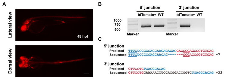
Figure 4. Evaluation of germline transmission of ErCas12a-mediated knockin with the NHEJ donor at the tbx2b locus. (A) Fluorescence reporter expression of F1 embryos obtained from outcross of a tbx2b NHEJ knockin F0 adult. Red fluorescence can be observed in the nervous system, eyes, pectoral fins, and endocardium of the F1 embryos. Scale bar: 200 μm. (B) Junction PCR results of the tbx2b knockin F1 embryos bearing red fluorescence. Primers tbx2b F1 and tbx2b R1 were used to amplify the 5′ junction, and primers bF2 and tbx2b R1 were used to amplify the 3′ junction. Note that due to the structure of the knockin allele, the 5′ junction amplicon of the of knockin allele is indistinguishable from that of the WT allele by electrophoresis. (C) Sequencing alignment of the junction PCR products after TA cloning. As the 5′ junction PCR product was a mixture of amplicons from the WT and knockin alleles, only sequencing results with indel mutations in the intronic junction site were considered to be amplified from the knockin allele. Blue characters indicate the tbx2b WT allele, red characters represent the donor sequences, and black characters represent random insertions resulting from NHEJ repair. Underlined characters indicate the PAMs of the tbx2b ErCas12a site and lamgolden Cas9 site for donor linearization.
Acknowledgments
This image is the copyrighted work of the attributed author or publisher, and
ZFIN has permission only to display this image to its users.
Additional permissions should be obtained from the applicable author or publisher of the image.
Full text @ Biology (Basel)

