IMAGE

Figure 1.

ID
ZDB-IMAGE-220328-12
Source
Figures for Krishnan et al., 2022
Image
Figure Caption

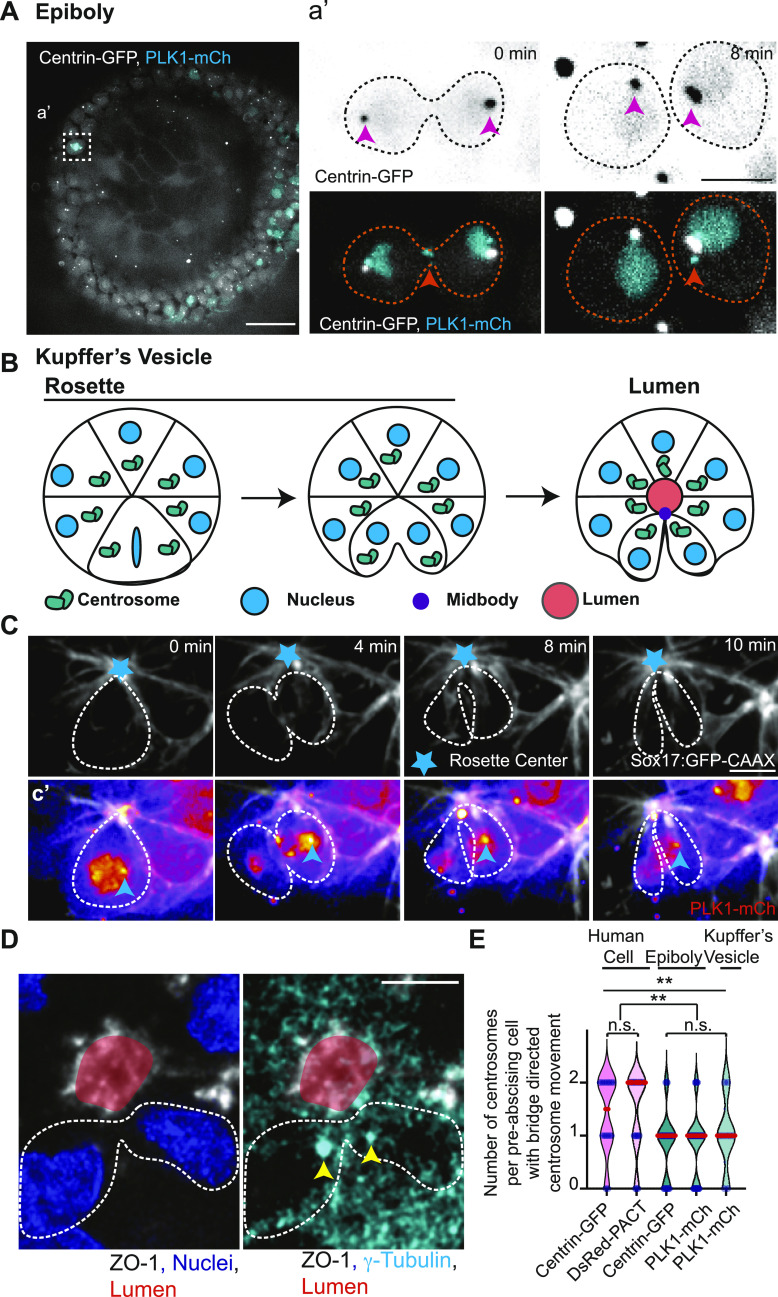
Figure 1. Differences in mitotic centrosome movement towards the cytokinetic bridge during pre-abscission between zebrafish embryos and human cells.

(A) Zebrafish embryo (5 h post fertilization) with centrin-GFP (gray) and PLK1-mCh (cyan). Scale bar, 50 μm. (a’) Inset of dividing cell in (A). Time-lapse of centrin-GFP (inverted grays, top panel; grays, bottom panel) and PLK1-mCh (cyan). Video 1 Pink arrow, centrosome. Orange arrow, midbody. Dashed lines, cell boundaries. Scale bar, 10 μm. (B) Model depicting centrosome (green) movement towards the cytokinetic bridge in dividing cells within the Kupffer’s vesicle (KV) during its development. Cyan, Nucleus. Purple, Midbody. Orange, Lumen. Dark lines, KV membranes. (C) Time-lapse of a dividing cell within the KV. KV cell membranes marked with Sox17:GFP-CAAX (gray). Cyan star, rosette center. Cyan arrow, centrosome. Dashed lines, cell boundaries. Scale Bar, 10 μm. (c’) Dividing cell depicted with PLK1-mCh (fire LUT). Cyan arrow, centrosome. (D) A KV pre-abscising cell fixed and immunostained for ZO-1 (gray), γ-tubulin (cyan) and DNA (DAPI, blue). Yellow arrow, centrosome. Dashed lines, cell boundaries. Scale bar, 10 μm. (E) Number of centrosomes per pre-abscising cell with bridge directed centrosome movement calculated as both centrosomes (2 centrosomes), only one centrosome (1 centrosome) and neither centrosome (0 centrosomes) moved shown as a violin plot with median (orange) and quartiles (dark dotted lines). Two-tailed t test between Centrin-GFP and DsRed-PACT in Human (HeLa) cells (pink background). n > 10 cells across n > 3 experiments n.s. not significant. One-way ANOVA across zebrafish epiboly cells and KV cells (green background), n.s. not significant. n > 10 cells across n > 2 embryos. One-way ANOVA, across all columns, **P < 0.01. Two-tailed t test between Human (HeLa) cells and zebrafish (Epiboly, KV) cells, **P < 0.01. n-values, detailed statistical results in Table S1.

Acknowledgments
This image is the copyrighted work of the attributed author or publisher, and ZFIN has permission only to display this image to its users. Additional permissions should be obtained from the applicable author or publisher of the image. Full text @ Life Sci Alliance