
Image
|
Figure Caption
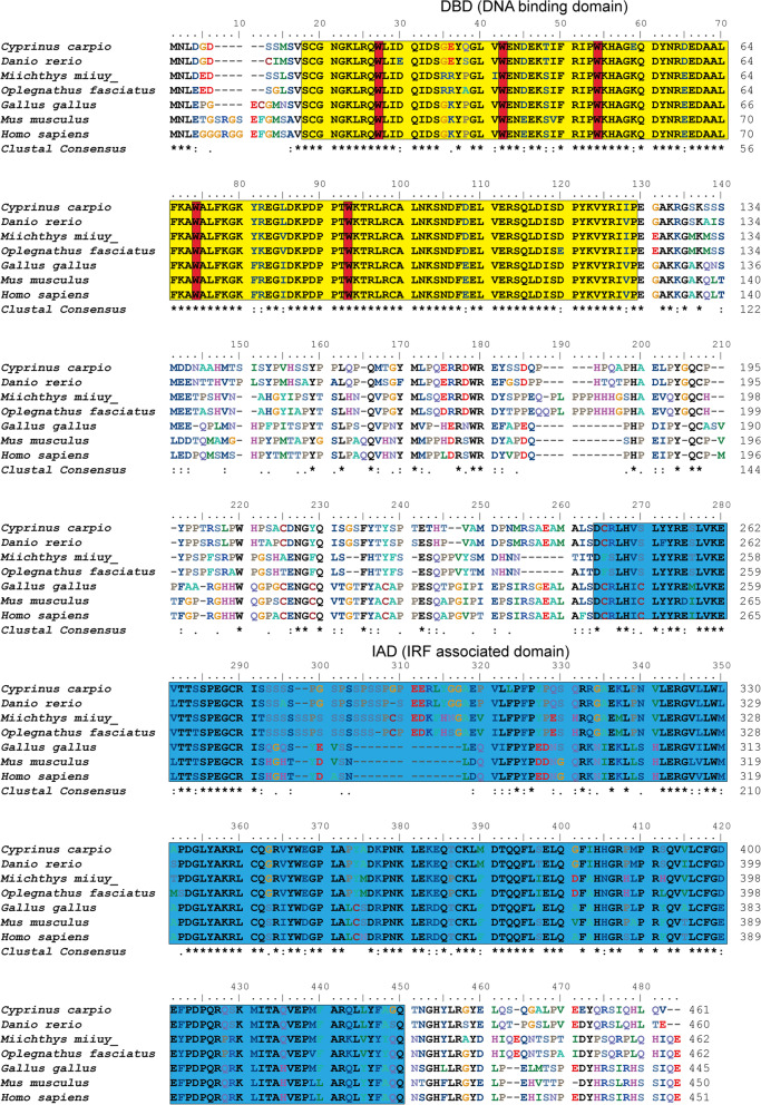
Fig. 1
Multiple alignment IRF4 protein sequences in different species. The sequences were aligned using the ClustalW method. Identical (*) and similar (: or .) residues are indicated; the predicted domains from SMART Server software have been indicated by colored boxes: the yellow denotes the DBD and the blue denotes the IAD. Five tryptophan (W) residues are boxed in red. The GenBank accession numbers of the genes are listed in Table
Acknowledgments
This image is the copyrighted work of the attributed author or publisher, and
ZFIN has permission only to display this image to its users.
Additional permissions should be obtained from the applicable author or publisher of the image.
Full text @ BMC Vet. Res.