
Image
|
Figure Caption
Figure 3
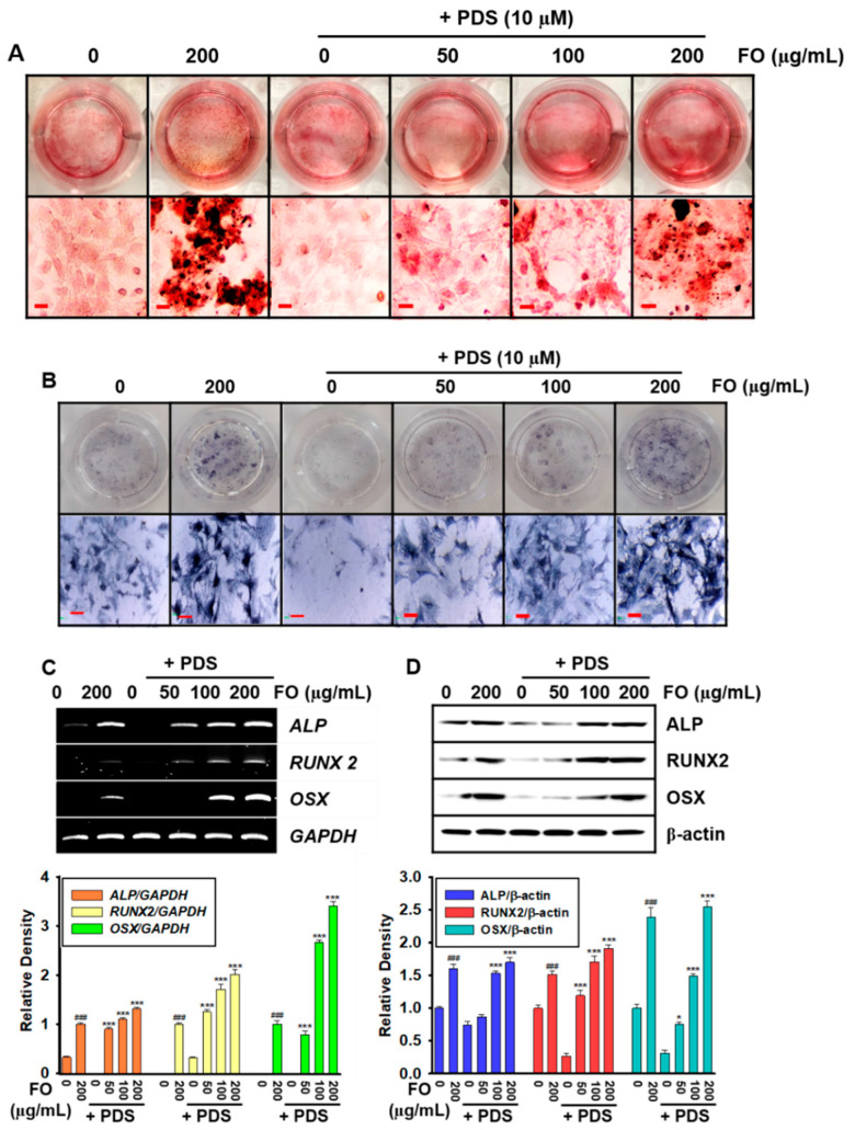
Figure 3. Prednisolone (PDS)-induced anti-osteogenic activity was inhibited by pretreatment with FO in MC3T3-E1 cells. MC3T3-E1 cells (1 × 10⁴ cells/mL) were pretreated with FO (0–200 μg/mL) for 2 h prior to treatment with 10 μM PDS for seven days. Fresh media with FO and/or PDS were replenished every two days. At day 7, (A) bone mineralization and (B) ALP activity were evaluated using alizarin red staining and a TRACP & ALP Double-Staining Kit, respectively. (C) Total mRNA were extracted, and RT-PCR was performed to evaluate the gene expressions of ALP, RUNX2, and OSX. GAPDH was used as the internal control. (D) Total proteins were extracted, and Western blotting was performed to evaluate the expression of ALP, RUNX2, and OSX. β-Actin was used as the internal control. All data are presented as means ± standard error of the mean (### p < 0.001 vs. untreated MC3T3-E1 cells; * p < 0.05, and *** p < 0.001 vs. PDS-treated MC3T3-E1 cells). ALP: alkaline phosphatase; RUNX2: runt-related transcription factor 2; and OSX: osterix.
Acknowledgments
This image is the copyrighted work of the attributed author or publisher, and
ZFIN has permission only to display this image to its users.
Additional permissions should be obtained from the applicable author or publisher of the image.
Full text @ Foods